sc-PDB
An Annotated Database of Druggable Binding Sites from the Protein DataBank
An Annotated Database of Druggable Binding Sites from the Protein DataBank
2.500 Å
X-ray
2013-02-25
| Name: | Dihydrolipoyl dehydrogenase |
|---|---|
| ID: | DLDH_ECOLI |
| AC: | P0A9P0 |
| Organism: | Escherichia coli |
| Reign: | Bacteria |
| TaxID: | 83333 |
| EC Number: | 1.8.1.4 |
| Chain Name: | Percentage of Residues within binding site |
|---|---|
| A | 95 % |
| B | 5 % |
| B-Factor: | 56.619 |
|---|---|
| Number of residues: | 70 |
| Including | |
| Standard Amino Acids: | 65 |
| Non Standard Amino Acids: | 0 |
| Water Molecules: | 5 |
| Cofactors: | |
| Metals: | |
| Ligandability | Volume (Å3) |
|---|---|
| 1.303 | 1417.500 |
| % Hydrophobic | % Polar |
|---|---|
| 51.43 | 48.57 |
| According to VolSite | |
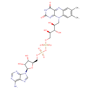
| HET Code: | FAD |
|---|---|
| Formula: | C27H31N9O15P2 |
| Molecular weight: | 783.534 g/mol |
| DrugBank ID: | DB03147 |
| Buried Surface Area: | 75.44 % |
| Polar Surface area: | 381.7 Å2 |
| Number of | |
|---|---|
| H-Bond Acceptors: | 22 |
| H-Bond Donors: | 7 |
| Rings: | 6 |
| Aromatic rings: | 3 |
| Anionic atoms: | 2 |
| Cationic atoms: | 0 |
| Rule of Five Violation: | 3 |
| Rotatable Bonds: | 13 |
| X | Y | Z |
|---|---|---|
| 36.4361 | 7.74257 | 48.9801 |


Represent the protein/ligand binding mode, centered on the ligand
Dashed lines represents hydrogen bonds and metal interactions
Green residue labels for amino acids with hydrophobic contacts (green lines) to the ligand
| Ligand | Protein | Interaction | |||
|---|---|---|---|---|---|
| Atom | Atom | Residue | Distance (Å) | Angle (°) | Type |
| C4' | CG | PRO- 16 | 4.3 | 0 | Hydrophobic |
| O1P | N | ALA- 17 | 3.14 | 136.44 | H-Bond (Protein Donor) |
| O3B | OE2 | GLU- 36 | 3.22 | 126.93 | H-Bond (Ligand Donor) |
| O3B | OE1 | GLU- 36 | 2.63 | 171.19 | H-Bond (Ligand Donor) |
| O2B | OE2 | GLU- 36 | 2.58 | 157.27 | H-Bond (Ligand Donor) |
| O2B | NE | ARG- 37 | 3.11 | 162.66 | H-Bond (Protein Donor) |
| N3A | N | ARG- 37 | 3.21 | 136.1 | H-Bond (Protein Donor) |
| C1B | CG | ARG- 37 | 4.36 | 0 | Hydrophobic |
| O1A | N | VAL- 44 | 2.97 | 141.38 | H-Bond (Protein Donor) |
| C8M | CG1 | VAL- 44 | 3.81 | 0 | Hydrophobic |
| C2' | CB | CYS- 45 | 4.41 | 0 | Hydrophobic |
| C4' | CB | CYS- 45 | 4.31 | 0 | Hydrophobic |
| O4' | N | CYS- 45 | 3.36 | 124.14 | H-Bond (Protein Donor) |
| O4 | NZ | LYS- 54 | 3.06 | 135.48 | H-Bond (Protein Donor) |
| N6A | O | GLY- 117 | 3.04 | 153.99 | H-Bond (Ligand Donor) |
| N1A | N | GLY- 117 | 2.97 | 169.32 | H-Bond (Protein Donor) |
| C7M | CB | SER- 165 | 3.7 | 0 | Hydrophobic |
| C8M | CB | SER- 165 | 4.43 | 0 | Hydrophobic |
| C7M | CD1 | LEU- 169 | 4.32 | 0 | Hydrophobic |
| C6 | CG1 | ILE- 186 | 4.05 | 0 | Hydrophobic |
| C7M | CG2 | ILE- 186 | 3.8 | 0 | Hydrophobic |
| C8 | CD1 | ILE- 186 | 3.63 | 0 | Hydrophobic |
| C8M | CD | ARG- 273 | 4.39 | 0 | Hydrophobic |
| O3' | OD2 | ASP- 313 | 3.23 | 139.45 | H-Bond (Ligand Donor) |
| O3' | OD1 | ASP- 313 | 2.9 | 158.14 | H-Bond (Ligand Donor) |
| C5' | CB | ASP- 313 | 4.4 | 0 | Hydrophobic |
| O2P | N | ASP- 313 | 3.01 | 155.81 | H-Bond (Protein Donor) |
| O2 | N | ALA- 321 | 2.71 | 157.27 | H-Bond (Protein Donor) |
| C2' | CB | ALA- 321 | 4.44 | 0 | Hydrophobic |
| C4' | CB | ALA- 321 | 4.41 | 0 | Hydrophobic |
| N3 | O | HIS- 445 | 2.78 | 123.01 | H-Bond (Ligand Donor) |
| O1P | O | HOH- 601 | 2.65 | 168.02 | H-Bond (Protein Donor) |
| O2P | O | HOH- 649 | 2.73 | 176.78 | H-Bond (Protein Donor) |
| O2A | O | HOH- 650 | 2.71 | 151.6 | H-Bond (Protein Donor) |
| N5 | O | HOH- 779 | 2.61 | 179.96 | H-Bond (Protein Donor) |
