sc-PDB
An Annotated Database of Druggable Binding Sites from the Protein DataBank
An Annotated Database of Druggable Binding Sites from the Protein DataBank
1.780 Å
X-ray
2012-12-28
| Name: | Methionine aminopeptidase 1 |
|---|---|
| ID: | MAP11_HUMAN |
| AC: | P53582 |
| Organism: | Homo sapiens |
| Reign: | Eukaryota |
| TaxID: | 9606 |
| EC Number: | / |
| Chain Name: | Percentage of Residues within binding site |
|---|---|
| A | 100 % |
| B-Factor: | 18.984 |
|---|---|
| Number of residues: | 23 |
| Including | |
| Standard Amino Acids: | 23 |
| Non Standard Amino Acids: | 0 |
| Water Molecules: | 0 |
| Cofactors: | |
| Metals: | |
| Ligandability | Volume (Å3) |
|---|---|
| 0.488 | 344.250 |
| % Hydrophobic | % Polar |
|---|---|
| 51.96 | 48.04 |
| According to VolSite | |
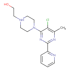
| HET Code: | PVP |
|---|---|
| Formula: | C16H21ClN5O |
| Molecular weight: | 334.824 g/mol |
| DrugBank ID: | - |
| Buried Surface Area: | 44.07 % |
| Polar Surface area: | 66.58 Å2 |
| Number of | |
|---|---|
| H-Bond Acceptors: | 5 |
| H-Bond Donors: | 2 |
| Rings: | 3 |
| Aromatic rings: | 2 |
| Anionic atoms: | 0 |
| Cationic atoms: | 1 |
| Rule of Five Violation: | 0 |
| Rotatable Bonds: | 4 |
| X | Y | Z |
|---|---|---|
| -2.29361 | -6.82461 | 15.792 |
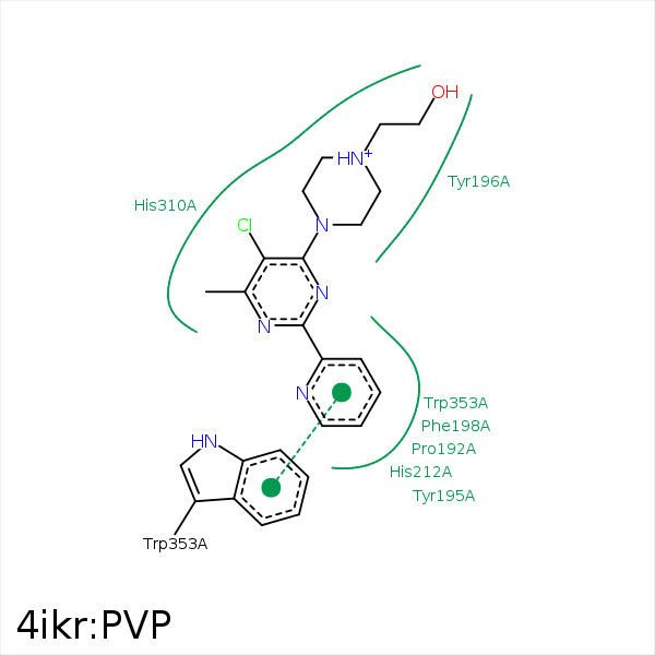


Represent the protein/ligand binding mode, centered on the ligand
Dashed lines represents hydrogen bonds and metal interactions
Green residue labels for amino acids with hydrophobic contacts (green lines) to the ligand
| Ligand | Protein | Interaction | |||
|---|---|---|---|---|---|
| Atom | Atom | Residue | Distance (Å) | Angle (°) | Type |
| C2 | CB | PRO- 192 | 4.05 | 0 | Hydrophobic |
| C13 | CE2 | TYR- 195 | 3.84 | 0 | Hydrophobic |
| C10 | CZ | TYR- 195 | 4.44 | 0 | Hydrophobic |
| C13 | CD1 | TYR- 196 | 4.18 | 0 | Hydrophobic |
| C3 | CB | PHE- 198 | 3.85 | 0 | Hydrophobic |
| C2 | SG | CYS- 203 | 3.8 | 0 | Hydrophobic |
| C10 | CB | HIS- 310 | 3.88 | 0 | Hydrophobic |
| CL | CB | HIS- 310 | 4.26 | 0 | Hydrophobic |
| DuAr | DuAr | HIS- 310 | 3.44 | 0 | Aromatic Face/Face |
| DuAr | DuAr | HIS- 310 | 3.44 | 0 | Aromatic Face/Face |
| C4 | CZ3 | TRP- 353 | 3.32 | 0 | Hydrophobic |
