sc-PDB
An Annotated Database of Druggable Binding Sites from the Protein DataBank
An Annotated Database of Druggable Binding Sites from the Protein DataBank
1.920 Å
X-ray
2012-10-22
| Name: | Glucose-1-phosphate thymidylyltransferase |
|---|---|
| ID: | Q9AGY4_ANETH |
| AC: | Q9AGY4 |
| Organism: | Aneurinibacillus thermoaerophilus |
| Reign: | Bacteria |
| TaxID: | 143495 |
| EC Number: | / |
| Chain Name: | Percentage of Residues within binding site |
|---|---|
| A | 100 % |
| B-Factor: | 26.654 |
|---|---|
| Number of residues: | 45 |
| Including | |
| Standard Amino Acids: | 42 |
| Non Standard Amino Acids: | 0 |
| Water Molecules: | 3 |
| Cofactors: | |
| Metals: | |
| Ligandability | Volume (Å3) |
|---|---|
| 0.010 | 472.500 |
| % Hydrophobic | % Polar |
|---|---|
| 47.14 | 52.86 |
| According to VolSite | |
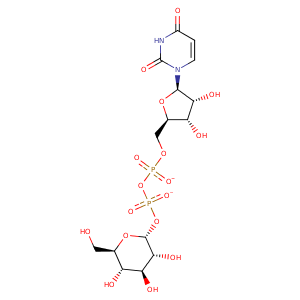
| HET Code: | UPG |
|---|---|
| Formula: | C15H22N2O17P2 |
| Molecular weight: | 564.286 g/mol |
| DrugBank ID: | DB01861 |
| Buried Surface Area: | 62.2 % |
| Polar Surface area: | 316.82 Å2 |
| Number of | |
|---|---|
| H-Bond Acceptors: | 17 |
| H-Bond Donors: | 7 |
| Rings: | 3 |
| Aromatic rings: | 0 |
| Anionic atoms: | 2 |
| Cationic atoms: | 0 |
| Rule of Five Violation: | 3 |
| Rotatable Bonds: | 9 |
| X | Y | Z |
|---|---|---|
| -2.49083 | -20.0823 | -17.2384 |
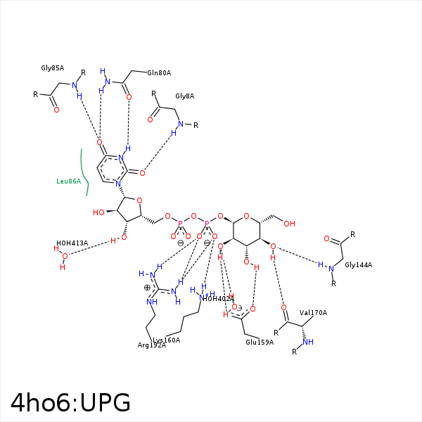


Represent the protein/ligand binding mode, centered on the ligand
Dashed lines represents hydrogen bonds and metal interactions
Green residue labels for amino acids with hydrophobic contacts (green lines) to the ligand
| Ligand | Protein | Interaction | |||
|---|---|---|---|---|---|
| Atom | Atom | Residue | Distance (Å) | Angle (°) | Type |
| O2 | N | GLY- 8 | 2.94 | 134.16 | H-Bond (Protein Donor) |
| O2C | O | GLY- 8 | 3.32 | 149.39 | H-Bond (Ligand Donor) |
| N3 | OE1 | GLN- 80 | 2.77 | 165.09 | H-Bond (Ligand Donor) |
| O4 | NE2 | GLN- 80 | 2.88 | 130.15 | H-Bond (Protein Donor) |
| O4 | N | GLY- 85 | 2.85 | 154.27 | H-Bond (Protein Donor) |
| C5C | CD1 | LEU- 86 | 4.2 | 0 | Hydrophobic |
| C2' | CD2 | LEU- 86 | 4.04 | 0 | Hydrophobic |
| C6' | CD1 | LEU- 106 | 4.25 | 0 | Hydrophobic |
| C5C | CD1 | LEU- 106 | 3.6 | 0 | Hydrophobic |
| C6' | CD1 | PHE- 143 | 4.16 | 0 | Hydrophobic |
| O3' | N | GLY- 144 | 3.17 | 123 | H-Bond (Protein Donor) |
| O4' | N | GLY- 144 | 3.01 | 145.14 | H-Bond (Protein Donor) |
| O2' | OE2 | GLU- 159 | 2.75 | 166.31 | H-Bond (Ligand Donor) |
| O3' | OE1 | GLU- 159 | 2.61 | 175.4 | H-Bond (Ligand Donor) |
| O3' | OE2 | GLU- 159 | 3.46 | 127.67 | H-Bond (Ligand Donor) |
| O2B | NZ | LYS- 160 | 3.02 | 131.08 | H-Bond (Protein Donor) |
| O2B | NZ | LYS- 160 | 3.02 | 0 | Ionic (Protein Cationic) |
| O4' | O | VAL- 170 | 2.68 | 175.04 | H-Bond (Ligand Donor) |
| O1B | CZ | ARG- 192 | 3.57 | 0 | Ionic (Protein Cationic) |
| O2B | CZ | ARG- 192 | 3.79 | 0 | Ionic (Protein Cationic) |
| O1B | NH1 | ARG- 192 | 3.12 | 146.68 | H-Bond (Protein Donor) |
| O1B | NH2 | ARG- 192 | 3.11 | 148.9 | H-Bond (Protein Donor) |
| O2B | NH2 | ARG- 192 | 2.96 | 133.31 | H-Bond (Protein Donor) |
| C2' | CG2 | ILE- 197 | 4.04 | 0 | Hydrophobic |
| C6' | CZ2 | TRP- 221 | 3.93 | 0 | Hydrophobic |
| O2' | O | HOH- 402 | 3.07 | 151.94 | H-Bond (Protein Donor) |
| O3C | O | HOH- 413 | 2.82 | 155.95 | H-Bond (Ligand Donor) |
