sc-PDB
An Annotated Database of Druggable Binding Sites from the Protein DataBank
An Annotated Database of Druggable Binding Sites from the Protein DataBank
1.330 Å
X-ray
2012-10-18
| Name: | Phenazine biosynthesis protein PhzG |
|---|---|
| ID: | PHZG_PSEFL |
| AC: | Q51793 |
| Organism: | Pseudomonas fluorescens |
| Reign: | Bacteria |
| TaxID: | 294 |
| EC Number: | 1.4 |
| Chain Name: | Percentage of Residues within binding site |
|---|---|
| A | 60 % |
| B | 40 % |
| B-Factor: | 8.488 |
|---|---|
| Number of residues: | 41 |
| Including | |
| Standard Amino Acids: | 36 |
| Non Standard Amino Acids: | 0 |
| Water Molecules: | 5 |
| Cofactors: | |
| Metals: | |
| Ligandability | Volume (Å3) |
|---|---|
| 1.023 | 1225.125 |
| % Hydrophobic | % Polar |
|---|---|
| 38.02 | 61.98 |
| According to VolSite | |
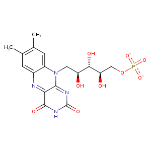
| HET Code: | FMN |
|---|---|
| Formula: | C17H19N4O9P |
| Molecular weight: | 454.328 g/mol |
| DrugBank ID: | DB03247 |
| Buried Surface Area: | 68.31 % |
| Polar Surface area: | 217.05 Å2 |
| Number of | |
|---|---|
| H-Bond Acceptors: | 12 |
| H-Bond Donors: | 4 |
| Rings: | 3 |
| Aromatic rings: | 1 |
| Anionic atoms: | 2 |
| Cationic atoms: | 0 |
| Rule of Five Violation: | 1 |
| Rotatable Bonds: | 7 |
| X | Y | Z |
|---|---|---|
| 12.8434 | 0.599806 | -4.16181 |
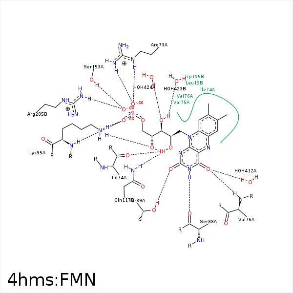


Represent the protein/ligand binding mode, centered on the ligand
Dashed lines represents hydrogen bonds and metal interactions
Green residue labels for amino acids with hydrophobic contacts (green lines) to the ligand
| Ligand | Protein | Interaction | |||
|---|---|---|---|---|---|
| Atom | Atom | Residue | Distance (Å) | Angle (°) | Type |
| C7M | CD1 | LEU- 19 | 3.52 | 0 | Hydrophobic |
| C8M | CD2 | LEU- 19 | 4.25 | 0 | Hydrophobic |
| C2' | CG | ARG- 73 | 4.01 | 0 | Hydrophobic |
| C3' | CB | ARG- 73 | 4.05 | 0 | Hydrophobic |
| O3P | CZ | ARG- 73 | 3.31 | 0 | Ionic (Protein Cationic) |
| O3P | NH1 | ARG- 73 | 2.9 | 143.94 | H-Bond (Protein Donor) |
| O3P | NE | ARG- 73 | 2.84 | 149.94 | H-Bond (Protein Donor) |
| O2' | O | ILE- 74 | 2.75 | 156.05 | H-Bond (Ligand Donor) |
| C7 | CG2 | ILE- 74 | 3.62 | 0 | Hydrophobic |
| C8 | CB | ILE- 74 | 4.04 | 0 | Hydrophobic |
| O4 | N | VAL- 76 | 3.09 | 176.39 | H-Bond (Protein Donor) |
| C6 | CG2 | VAL- 76 | 3.78 | 0 | Hydrophobic |
| N3 | O | SER- 88 | 2.87 | 167.79 | H-Bond (Ligand Donor) |
| O2 | OG1 | THR- 89 | 2.76 | 165.63 | H-Bond (Protein Donor) |
| O4' | NZ | LYS- 95 | 2.75 | 156.05 | H-Bond (Protein Donor) |
| O1P | NZ | LYS- 95 | 2.8 | 152.75 | H-Bond (Protein Donor) |
| O1P | N | LYS- 95 | 2.85 | 165.33 | H-Bond (Protein Donor) |
| O1P | NZ | LYS- 95 | 2.8 | 0 | Ionic (Protein Cationic) |
| C7M | CZ | TYR- 110 | 3.38 | 0 | Hydrophobic |
| C8M | CE1 | TYR- 110 | 3.83 | 0 | Hydrophobic |
| C8M | CG | GLN- 117 | 3.7 | 0 | Hydrophobic |
| O2' | NE2 | GLN- 117 | 2.94 | 136.02 | H-Bond (Protein Donor) |
| O2 | NE2 | GLN- 152 | 3.42 | 152.42 | H-Bond (Protein Donor) |
| C5' | CG | GLN- 152 | 3.96 | 0 | Hydrophobic |
| O2P | OG | SER- 153 | 2.58 | 159.08 | H-Bond (Protein Donor) |
| C8M | CZ3 | TRP- 195 | 3.62 | 0 | Hydrophobic |
| O2P | NH2 | ARG- 205 | 2.98 | 161.31 | H-Bond (Protein Donor) |
| O2P | CZ | ARG- 205 | 3.91 | 0 | Ionic (Protein Cationic) |
| O4 | O | HOH- 412 | 2.8 | 179.97 | H-Bond (Protein Donor) |
| O3' | O | HOH- 423 | 2.92 | 152.08 | H-Bond (Ligand Donor) |
| O3' | O | HOH- 424 | 2.62 | 179.97 | H-Bond (Protein Donor) |
