sc-PDB
An Annotated Database of Druggable Binding Sites from the Protein DataBank
An Annotated Database of Druggable Binding Sites from the Protein DataBank
2.750 Å
X-ray
2012-10-09
| Name: | Phototropin-1 |
|---|---|
| ID: | PHOT1_ARATH |
| AC: | O48963 |
| Organism: | Arabidopsis thaliana |
| Reign: | Eukaryota |
| TaxID: | 3702 |
| EC Number: | 2.7.11.1 |
| Chain Name: | Percentage of Residues within binding site |
|---|---|
| A | 100 % |
| B-Factor: | 43.033 |
|---|---|
| Number of residues: | 31 |
| Including | |
| Standard Amino Acids: | 31 |
| Non Standard Amino Acids: | 0 |
| Water Molecules: | 0 |
| Cofactors: | |
| Metals: | |
| Ligandability | Volume (Å3) |
|---|---|
| 0.635 | 411.750 |
| % Hydrophobic | % Polar |
|---|---|
| 55.74 | 44.26 |
| According to VolSite | |
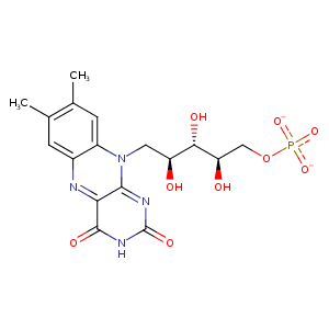
| HET Code: | FMN |
|---|---|
| Formula: | C17H19N4O9P |
| Molecular weight: | 454.328 g/mol |
| DrugBank ID: | DB03247 |
| Buried Surface Area: | 71.12 % |
| Polar Surface area: | 217.05 Å2 |
| Number of | |
|---|---|
| H-Bond Acceptors: | 12 |
| H-Bond Donors: | 4 |
| Rings: | 3 |
| Aromatic rings: | 1 |
| Anionic atoms: | 2 |
| Cationic atoms: | 0 |
| Rule of Five Violation: | 1 |
| Rotatable Bonds: | 7 |
| X | Y | Z |
|---|---|---|
| 12.2507 | -7.87626 | 11.5273 |
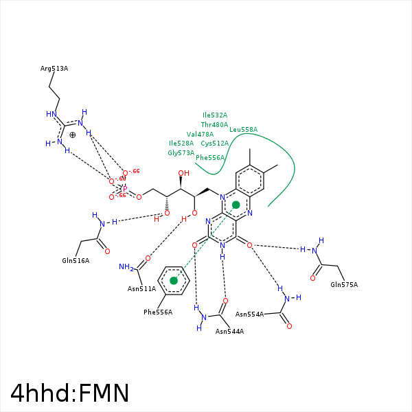


Represent the protein/ligand binding mode, centered on the ligand
Dashed lines represents hydrogen bonds and metal interactions
Green residue labels for amino acids with hydrophobic contacts (green lines) to the ligand
| Ligand | Protein | Interaction | |||
|---|---|---|---|---|---|
| Atom | Atom | Residue | Distance (Å) | Angle (°) | Type |
| C6 | CG2 | VAL- 478 | 3.6 | 0 | Hydrophobic |
| C8M | CB | THR- 480 | 4.16 | 0 | Hydrophobic |
| C7M | CG2 | THR- 480 | 3.56 | 0 | Hydrophobic |
| O2' | OD1 | ASN- 511 | 2.56 | 156.76 | H-Bond (Ligand Donor) |
| C2' | CB | CYS- 512 | 4.32 | 0 | Hydrophobic |
| C6 | SG | CYS- 512 | 3.79 | 0 | Hydrophobic |
| C9A | CB | CYS- 512 | 3.77 | 0 | Hydrophobic |
| O1P | NH1 | ARG- 513 | 2.62 | 157.84 | H-Bond (Protein Donor) |
| O1P | NH2 | ARG- 513 | 3.06 | 132.22 | H-Bond (Protein Donor) |
| O2P | NH1 | ARG- 513 | 2.92 | 123.68 | H-Bond (Protein Donor) |
| O1P | CZ | ARG- 513 | 3.26 | 0 | Ionic (Protein Cationic) |
| O2 | NE2 | GLN- 516 | 3.32 | 150.76 | H-Bond (Protein Donor) |
| O4' | NE2 | GLN- 516 | 3.11 | 142.55 | H-Bond (Protein Donor) |
| C5' | CG1 | VAL- 525 | 3.7 | 0 | Hydrophobic |
| C1' | CG2 | ILE- 528 | 3.75 | 0 | Hydrophobic |
| C5' | CG | ARG- 529 | 3.68 | 0 | Hydrophobic |
| O3P | CZ | ARG- 529 | 3.44 | 0 | Ionic (Protein Cationic) |
| C8M | CD1 | ILE- 532 | 4.19 | 0 | Hydrophobic |
| C9 | CD1 | ILE- 532 | 4.29 | 0 | Hydrophobic |
| O2 | ND2 | ASN- 544 | 2.86 | 136.41 | H-Bond (Protein Donor) |
| N3 | OD1 | ASN- 544 | 2.81 | 159.78 | H-Bond (Ligand Donor) |
| O4 | ND2 | ASN- 554 | 3.18 | 140.59 | H-Bond (Protein Donor) |
| C7 | CD1 | LEU- 558 | 4.19 | 0 | Hydrophobic |
| C8M | CD1 | LEU- 558 | 3.78 | 0 | Hydrophobic |
| C7M | CB | PHE- 571 | 3.98 | 0 | Hydrophobic |
| C8M | CB | PHE- 571 | 3.76 | 0 | Hydrophobic |
| O4 | NE2 | GLN- 575 | 2.9 | 137.99 | H-Bond (Protein Donor) |
| N5 | NE2 | GLN- 575 | 3.47 | 138.78 | H-Bond (Protein Donor) |
