sc-PDB
An Annotated Database of Druggable Binding Sites from the Protein DataBank
An Annotated Database of Druggable Binding Sites from the Protein DataBank
2.130 Å
X-ray
2012-07-01
| Name: | Lysine-specific histone demethylase 1B |
|---|---|
| ID: | KDM1B_HUMAN |
| AC: | Q8NB78 |
| Organism: | Homo sapiens |
| Reign: | Eukaryota |
| TaxID: | 9606 |
| EC Number: | 1 |
| Chain Name: | Percentage of Residues within binding site |
|---|---|
| A | 100 % |
| B-Factor: | 38.854 |
|---|---|
| Number of residues: | 69 |
| Including | |
| Standard Amino Acids: | 64 |
| Non Standard Amino Acids: | 0 |
| Water Molecules: | 5 |
| Cofactors: | |
| Metals: | |
| Ligandability | Volume (Å3) |
|---|---|
| 1.237 | 570.375 |
| % Hydrophobic | % Polar |
|---|---|
| 57.40 | 42.60 |
| According to VolSite | |
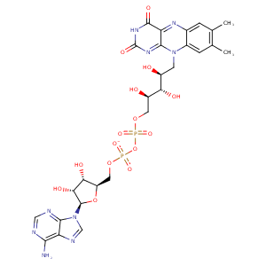
| HET Code: | FAD |
|---|---|
| Formula: | C27H31N9O15P2 |
| Molecular weight: | 783.534 g/mol |
| DrugBank ID: | DB03147 |
| Buried Surface Area: | 83.81 % |
| Polar Surface area: | 381.7 Å2 |
| Number of | |
|---|---|
| H-Bond Acceptors: | 22 |
| H-Bond Donors: | 7 |
| Rings: | 6 |
| Aromatic rings: | 3 |
| Anionic atoms: | 2 |
| Cationic atoms: | 0 |
| Rule of Five Violation: | 3 |
| Rotatable Bonds: | 13 |
| X | Y | Z |
|---|---|---|
| -29.1631 | -1.98696 | -49.3689 |
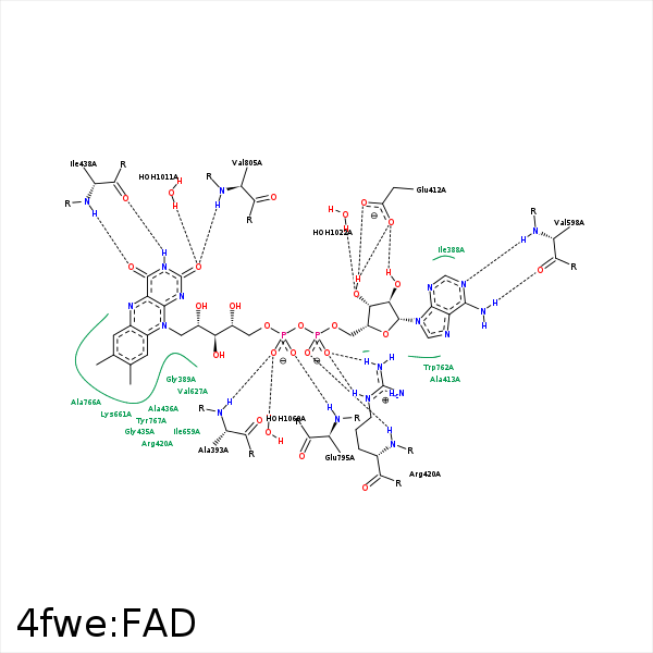


Represent the protein/ligand binding mode, centered on the ligand
Dashed lines represents hydrogen bonds and metal interactions
Green residue labels for amino acids with hydrophobic contacts (green lines) to the ligand
| Ligand | Protein | Interaction | |||
|---|---|---|---|---|---|
| Atom | Atom | Residue | Distance (Å) | Angle (°) | Type |
| C4' | CG | PRO- 392 | 4.26 | 0 | Hydrophobic |
| O1P | N | ALA- 393 | 2.87 | 163.64 | H-Bond (Protein Donor) |
| O3B | OE1 | GLU- 412 | 2.73 | 163.19 | H-Bond (Ligand Donor) |
| O3B | OE2 | GLU- 412 | 3.1 | 130.07 | H-Bond (Ligand Donor) |
| O2B | OE2 | GLU- 412 | 2.52 | 159.08 | H-Bond (Ligand Donor) |
| N3A | N | ALA- 413 | 3.24 | 138.67 | H-Bond (Protein Donor) |
| O1A | N | ARG- 420 | 2.86 | 173.63 | H-Bond (Protein Donor) |
| O2A | NE | ARG- 420 | 2.68 | 160.29 | H-Bond (Protein Donor) |
| O2A | NH2 | ARG- 420 | 3.05 | 135.21 | H-Bond (Protein Donor) |
| O3P | NH2 | ARG- 420 | 3.22 | 129.25 | H-Bond (Protein Donor) |
| O2A | CZ | ARG- 420 | 3.29 | 0 | Ionic (Protein Cationic) |
| C8M | CG | ARG- 420 | 3.89 | 0 | Hydrophobic |
| C9 | CB | ARG- 420 | 4.19 | 0 | Hydrophobic |
| C3' | CB | ARG- 420 | 4.11 | 0 | Hydrophobic |
| C9A | CB | ALA- 436 | 4.1 | 0 | Hydrophobic |
| C2' | CB | ALA- 436 | 4.07 | 0 | Hydrophobic |
| O4 | N | GLN- 437 | 3.43 | 160.95 | H-Bond (Protein Donor) |
| N3 | O | ILE- 438 | 2.78 | 152.26 | H-Bond (Ligand Donor) |
| O4 | N | ILE- 438 | 2.95 | 146.99 | H-Bond (Protein Donor) |
| N6A | O | VAL- 598 | 2.93 | 160.4 | H-Bond (Ligand Donor) |
| N1A | N | VAL- 598 | 2.87 | 165.83 | H-Bond (Protein Donor) |
| C5B | CG | PRO- 628 | 4.14 | 0 | Hydrophobic |
| C7M | CG1 | ILE- 659 | 3.88 | 0 | Hydrophobic |
| C6 | CD1 | ILE- 659 | 3.84 | 0 | Hydrophobic |
| C7M | CG | LYS- 661 | 4 | 0 | Hydrophobic |
| C7M | CE2 | TRP- 757 | 4.38 | 0 | Hydrophobic |
| C8M | CD2 | TRP- 757 | 3.81 | 0 | Hydrophobic |
| C2B | CB | TRP- 762 | 4.28 | 0 | Hydrophobic |
| C3B | CD1 | ILE- 763 | 3.63 | 0 | Hydrophobic |
| C8M | CB | ALA- 766 | 3.44 | 0 | Hydrophobic |
| C1' | CD2 | TYR- 767 | 3.76 | 0 | Hydrophobic |
| C3' | CG | GLU- 795 | 4.41 | 0 | Hydrophobic |
| C5' | CB | GLU- 795 | 3.72 | 0 | Hydrophobic |
| O2P | N | GLU- 795 | 2.86 | 162.69 | H-Bond (Protein Donor) |
| O3' | O | THR- 804 | 3.47 | 122.53 | H-Bond (Ligand Donor) |
| O2 | N | VAL- 805 | 2.82 | 160.91 | H-Bond (Protein Donor) |
| C2' | CG2 | VAL- 805 | 3.9 | 0 | Hydrophobic |
| C4' | CG2 | VAL- 805 | 4.3 | 0 | Hydrophobic |
| C5' | CB | ALA- 808 | 3.72 | 0 | Hydrophobic |
| O2 | O | HOH- 1011 | 2.62 | 163.55 | H-Bond (Protein Donor) |
| O3B | O | HOH- 1022 | 2.82 | 179.98 | H-Bond (Protein Donor) |
| O2 | O | HOH- 1023 | 3.49 | 150.83 | H-Bond (Protein Donor) |
| O1P | O | HOH- 1060 | 2.7 | 159.92 | H-Bond (Protein Donor) |
