sc-PDB
An Annotated Database of Druggable Binding Sites from the Protein DataBank
An Annotated Database of Druggable Binding Sites from the Protein DataBank
1.500 Å
X-ray
2012-04-19
| Name: | Coenzyme A disulfide reductase |
|---|---|
| ID: | CDR_STAA3 |
| AC: | Q2FIA5 |
| Organism: | Staphylococcus aureus |
| Reign: | Bacteria |
| TaxID: | 367830 |
| EC Number: | / |
| Chain Name: | Percentage of Residues within binding site |
|---|---|
| A | 92 % |
| B | 8 % |
| B-Factor: | 16.364 |
|---|---|
| Number of residues: | 64 |
| Including | |
| Standard Amino Acids: | 58 |
| Non Standard Amino Acids: | 1 |
| Water Molecules: | 5 |
| Cofactors: | COA |
| Metals: | |
| Ligandability | Volume (Å3) |
|---|---|
| 0.603 | 1836.000 |
| % Hydrophobic | % Polar |
|---|---|
| 48.71 | 51.29 |
| According to VolSite | |
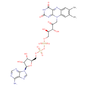
| HET Code: | FAD |
|---|---|
| Formula: | C27H31N9O15P2 |
| Molecular weight: | 783.534 g/mol |
| DrugBank ID: | DB03147 |
| Buried Surface Area: | 77.55 % |
| Polar Surface area: | 381.7 Å2 |
| Number of | |
|---|---|
| H-Bond Acceptors: | 22 |
| H-Bond Donors: | 7 |
| Rings: | 6 |
| Aromatic rings: | 3 |
| Anionic atoms: | 2 |
| Cationic atoms: | 0 |
| Rule of Five Violation: | 3 |
| Rotatable Bonds: | 13 |
| X | Y | Z |
|---|---|---|
| 23.5155 | 10.7181 | -11.8549 |


Represent the protein/ligand binding mode, centered on the ligand
Dashed lines represents hydrogen bonds and metal interactions
Green residue labels for amino acids with hydrophobic contacts (green lines) to the ligand
| Ligand | Protein | Interaction | |||
|---|---|---|---|---|---|
| Atom | Atom | Residue | Distance (Å) | Angle (°) | Type |
| C4B | CG2 | VAL- 10 | 4.04 | 0 | Hydrophobic |
| O2A | N | ALA- 11 | 3.24 | 163.3 | H-Bond (Protein Donor) |
| C4' | CB | ALA- 11 | 3.57 | 0 | Hydrophobic |
| O1P | N | GLY- 12 | 2.82 | 152.63 | H-Bond (Protein Donor) |
| O3B | OE1 | GLU- 33 | 2.71 | 170.78 | H-Bond (Ligand Donor) |
| O3B | OE2 | GLU- 33 | 3.06 | 126.38 | H-Bond (Ligand Donor) |
| O2B | OE2 | GLU- 33 | 2.75 | 145.63 | H-Bond (Ligand Donor) |
| O2B | NZ | LYS- 34 | 3.12 | 141.51 | H-Bond (Protein Donor) |
| N3A | N | LYS- 34 | 3.07 | 142.54 | H-Bond (Protein Donor) |
| C2B | CE | LYS- 34 | 3.89 | 0 | Hydrophobic |
| O2A | ND2 | ASN- 42 | 2.94 | 139.03 | H-Bond (Protein Donor) |
| C9 | CB | ASN- 42 | 4.09 | 0 | Hydrophobic |
| C9A | SG | CYS- 43 | 3.85 | 0 | Hydrophobic |
| C7M | CB | LEU- 45 | 4.33 | 0 | Hydrophobic |
| C6 | CG | PRO- 46 | 4.37 | 0 | Hydrophobic |
| C7M | CG | PRO- 46 | 4 | 0 | Hydrophobic |
| N6A | O | VAL- 81 | 2.79 | 164.84 | H-Bond (Ligand Donor) |
| N1A | N | VAL- 81 | 2.9 | 155.64 | H-Bond (Protein Donor) |
| O1P | OG | SER- 112 | 2.66 | 144.71 | H-Bond (Protein Donor) |
| C7M | CD2 | LEU- 130 | 3.61 | 0 | Hydrophobic |
| C8M | CD | ARG- 131 | 3.91 | 0 | Hydrophobic |
| C6 | CG2 | VAL- 159 | 3.75 | 0 | Hydrophobic |
| C9A | CG2 | VAL- 159 | 4.48 | 0 | Hydrophobic |
| C7M | CG1 | VAL- 159 | 3.45 | 0 | Hydrophobic |
| O3' | OD1 | ASP- 277 | 2.86 | 174.41 | H-Bond (Ligand Donor) |
| O2P | N | ASP- 277 | 2.88 | 156.49 | H-Bond (Protein Donor) |
| N1 | N | ALA- 295 | 3.47 | 136.67 | H-Bond (Protein Donor) |
| O2 | N | ALA- 295 | 2.8 | 164 | H-Bond (Protein Donor) |
| C5' | CB | ALA- 298 | 4.07 | 0 | Hydrophobic |
| N3 | O | PHE- 419 | 2.96 | 169.45 | H-Bond (Ligand Donor) |
| O1P | O | HOH- 601 | 2.71 | 134.44 | H-Bond (Protein Donor) |
| O2P | O | HOH- 604 | 2.7 | 179.96 | H-Bond (Protein Donor) |
| O3B | O | HOH- 611 | 2.76 | 179.98 | H-Bond (Protein Donor) |
| O1A | O | HOH- 614 | 2.87 | 139.57 | H-Bond (Protein Donor) |
