sc-PDB
An Annotated Database of Druggable Binding Sites from the Protein DataBank
An Annotated Database of Druggable Binding Sites from the Protein DataBank
2.950 Å
X-ray
2012-01-12
| Name: | Purine nucleoside phosphorylase DeoD-type |
|---|---|
| ID: | DEOD_BACSU |
| AC: | O34925 |
| Organism: | Bacillus subtilis |
| Reign: | Bacteria |
| TaxID: | 224308 |
| EC Number: | 2.4.2.1 |
| Chain Name: | Percentage of Residues within binding site |
|---|---|
| A | 100 % |
| B-Factor: | 58.467 |
|---|---|
| Number of residues: | 27 |
| Including | |
| Standard Amino Acids: | 26 |
| Non Standard Amino Acids: | 1 |
| Water Molecules: | 0 |
| Cofactors: | |
| Metals: | CL |
| Ligandability | Volume (Å3) |
|---|---|
| 0.829 | 580.500 |
| % Hydrophobic | % Polar |
|---|---|
| 50.58 | 49.42 |
| According to VolSite | |
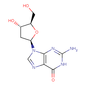
| HET Code: | GNG |
|---|---|
| Formula: | C10H13N5O4 |
| Molecular weight: | 267.241 g/mol |
| DrugBank ID: | - |
| Buried Surface Area: | 59.95 % |
| Polar Surface area: | 134.99 Å2 |
| Number of | |
|---|---|
| H-Bond Acceptors: | 8 |
| H-Bond Donors: | 4 |
| Rings: | 3 |
| Aromatic rings: | 1 |
| Anionic atoms: | 0 |
| Cationic atoms: | 0 |
| Rule of Five Violation: | 0 |
| Rotatable Bonds: | 2 |
| X | Y | Z |
|---|---|---|
| -7.15326 | -54.2366 | -23.5374 |
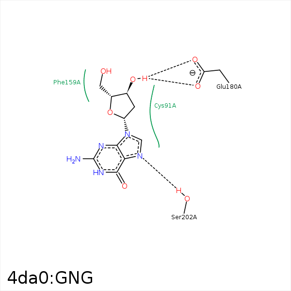


Represent the protein/ligand binding mode, centered on the ligand
Dashed lines represents hydrogen bonds and metal interactions
Green residue labels for amino acids with hydrophobic contacts (green lines) to the ligand
| Ligand | Protein | Interaction | |||
|---|---|---|---|---|---|
| Atom | Atom | Residue | Distance (Å) | Angle (°) | Type |
| C5' | SD | MET- 64 | 3.99 | 0 | Hydrophobic |
| C5' | CE1 | PHE- 159 | 3.6 | 0 | Hydrophobic |
| C2' | CB | GLU- 178 | 3.7 | 0 | Hydrophobic |
| C2' | CG | MET- 179 | 3.91 | 0 | Hydrophobic |
| C3' | SD | MET- 179 | 3.65 | 0 | Hydrophobic |
| O3' | OE1 | GLU- 180 | 2.64 | 136.16 | H-Bond (Ligand Donor) |
| O3' | OE2 | GLU- 180 | 2.61 | 134.65 | H-Bond (Ligand Donor) |
| N7 | OG | SER- 202 | 2.69 | 156.61 | H-Bond (Protein Donor) |
