sc-PDB
An Annotated Database of Druggable Binding Sites from the Protein DataBank
An Annotated Database of Druggable Binding Sites from the Protein DataBank
1.750 Å
X-ray
2013-11-11
| Name: | Integrase |
|---|---|
| ID: | Q76353_9HIV1 |
| AC: | Q76353 |
| Organism: | Human immunodeficiency virus 1 |
| Reign: | Viruses |
| TaxID: | 11676 |
| EC Number: | / |
| Chain Name: | Percentage of Residues within binding site |
|---|---|
| A | 61 % |
| B | 39 % |
| B-Factor: | 15.992 |
|---|---|
| Number of residues: | 28 |
| Including | |
| Standard Amino Acids: | 28 |
| Non Standard Amino Acids: | 0 |
| Water Molecules: | 0 |
| Cofactors: | |
| Metals: | |
| Ligandability | Volume (Å3) |
|---|---|
| 0.646 | 381.375 |
| % Hydrophobic | % Polar |
|---|---|
| 47.79 | 52.21 |
| According to VolSite | |
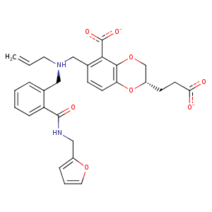
| HET Code: | 9NS |
|---|---|
| Formula: | C29H29N2O8 |
| Molecular weight: | 533.549 g/mol |
| DrugBank ID: | - |
| Buried Surface Area: | 51.82 % |
| Polar Surface area: | 145.4 Å2 |
| Number of | |
|---|---|
| H-Bond Acceptors: | 7 |
| H-Bond Donors: | 2 |
| Rings: | 4 |
| Aromatic rings: | 3 |
| Anionic atoms: | 2 |
| Cationic atoms: | 1 |
| Rule of Five Violation: | 1 |
| Rotatable Bonds: | 13 |
| X | Y | Z |
|---|---|---|
| 33.6187 | -7.72169 | 13.65 |
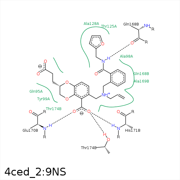


Represent the protein/ligand binding mode, centered on the ligand
Dashed lines represents hydrogen bonds and metal interactions
Green residue labels for amino acids with hydrophobic contacts (green lines) to the ligand
| Ligand | Protein | Interaction | |||
|---|---|---|---|---|---|
| Atom | Atom | Residue | Distance (Å) | Angle (°) | Type |
| C7 | CG | GLN- 95 | 4.1 | 0 | Hydrophobic |
| C28 | CG | GLU- 96 | 4.48 | 0 | Hydrophobic |
| C7 | CB | ALA- 98 | 4.43 | 0 | Hydrophobic |
| C22 | CD1 | TYR- 99 | 4.3 | 0 | Hydrophobic |
| C23 | CB | TYR- 99 | 4.24 | 0 | Hydrophobic |
| C28 | CB | TYR- 99 | 3.78 | 0 | Hydrophobic |
| C29 | CG | TYR- 99 | 3.9 | 0 | Hydrophobic |
| C14 | CB | TYR- 99 | 4.2 | 0 | Hydrophobic |
| C6 | CG2 | THR- 125 | 4.25 | 0 | Hydrophobic |
| N30 | O | GLN- 168 | 2.74 | 168.39 | H-Bond (Ligand Donor) |
| O35 | N | GLU- 170 | 2.84 | 161.88 | H-Bond (Protein Donor) |
| C22 | CB | HIS- 171 | 4.16 | 0 | Hydrophobic |
| O32 | N | HIS- 171 | 2.92 | 165.12 | H-Bond (Protein Donor) |
| C22 | CG | LYS- 173 | 3.93 | 0 | Hydrophobic |
| C29 | CD | LYS- 173 | 4.25 | 0 | Hydrophobic |
| C15 | CB | THR- 174 | 4.22 | 0 | Hydrophobic |
| C11 | CG2 | THR- 174 | 4.07 | 0 | Hydrophobic |
| O32 | OG1 | THR- 174 | 2.74 | 163.66 | H-Bond (Protein Donor) |
