sc-PDB
An Annotated Database of Druggable Binding Sites from the Protein DataBank
An Annotated Database of Druggable Binding Sites from the Protein DataBank
2.600 Å
X-ray
2013-05-04
| Name: | NTPase P4 |
|---|---|
| ID: | Q94M05_9VIRU |
| AC: | Q94M05 |
| Organism: | Pseudomonas phage phi12 |
| Reign: | Viruses |
| TaxID: | 161736 |
| EC Number: | / |
| Chain Name: | Percentage of Residues within binding site |
|---|---|
| B | 43 % |
| C | 57 % |
| B-Factor: | 61.401 |
|---|---|
| Number of residues: | 38 |
| Including | |
| Standard Amino Acids: | 38 |
| Non Standard Amino Acids: | 0 |
| Water Molecules: | 0 |
| Cofactors: | |
| Metals: | |
| Ligandability | Volume (Å3) |
|---|---|
| 0.344 | 725.625 |
| % Hydrophobic | % Polar |
|---|---|
| 33.95 | 66.05 |
| According to VolSite | |
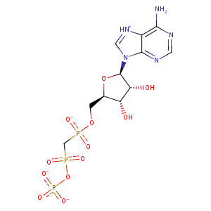
| HET Code: | APC |
|---|---|
| Formula: | C11H14N5O12P3 |
| Molecular weight: | 501.176 g/mol |
| DrugBank ID: | DB02596 |
| Buried Surface Area: | 50.04 % |
| Polar Surface area: | 310.64 Å2 |
| Number of | |
|---|---|
| H-Bond Acceptors: | 16 |
| H-Bond Donors: | 3 |
| Rings: | 3 |
| Aromatic rings: | 2 |
| Anionic atoms: | 4 |
| Cationic atoms: | 0 |
| Rule of Five Violation: | 2 |
| Rotatable Bonds: | 8 |
| X | Y | Z |
|---|---|---|
| 32.3348 | 53.3705 | 27.198 |
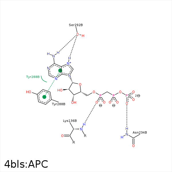


Represent the protein/ligand binding mode, centered on the ligand
Dashed lines represents hydrogen bonds and metal interactions
Green residue labels for amino acids with hydrophobic contacts (green lines) to the ligand
| Ligand | Protein | Interaction | |||
|---|---|---|---|---|---|
| Atom | Atom | Residue | Distance (Å) | Angle (°) | Type |
| C1' | CG | PRO- 138 | 4.46 | 0 | Hydrophobic |
| O1G | NZ | LYS- 192 | 3.51 | 0 | Ionic (Protein Cationic) |
| O2G | NZ | LYS- 192 | 2.86 | 0 | Ionic (Protein Cationic) |
| O3G | CZ | ARG- 272 | 3.67 | 0 | Ionic (Protein Cationic) |
| C1' | CZ | TYR- 288 | 4.34 | 0 | Hydrophobic |
| DuAr | DuAr | TYR- 288 | 3.69 | 0 | Aromatic Face/Face |
| N6 | OG | SER- 292 | 3.14 | 165.31 | H-Bond (Ligand Donor) |
