sc-PDB
An Annotated Database of Druggable Binding Sites from the Protein DataBank
An Annotated Database of Druggable Binding Sites from the Protein DataBank
2.100 Å
X-ray
2012-07-30
| Name: | Glucose-1-phosphate thymidylyltransferase |
|---|---|
| ID: | Q9HU22_PSEAE |
| AC: | Q9HU22 |
| Organism: | Pseudomonas aeruginosa |
| Reign: | Bacteria |
| TaxID: | 208964 |
| EC Number: | / |
| Chain Name: | Percentage of Residues within binding site |
|---|---|
| D | 100 % |
| B-Factor: | 70.008 |
|---|---|
| Number of residues: | 20 |
| Including | |
| Standard Amino Acids: | 18 |
| Non Standard Amino Acids: | 1 |
| Water Molecules: | 1 |
| Cofactors: | |
| Metals: | CL |
| Ligandability | Volume (Å3) |
|---|---|
| 1.105 | 560.250 |
| % Hydrophobic | % Polar |
|---|---|
| 47.59 | 52.41 |
| According to VolSite | |
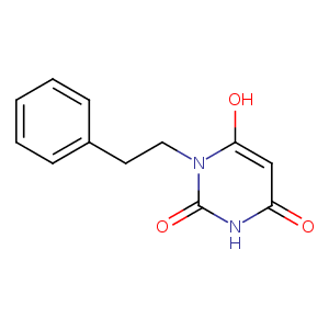
| HET Code: | GJB |
|---|---|
| Formula: | C12H11N2O3 |
| Molecular weight: | 231.227 g/mol |
| DrugBank ID: | - |
| Buried Surface Area: | 34.85 % |
| Polar Surface area: | 86.31 Å2 |
| Number of | |
|---|---|
| H-Bond Acceptors: | 3 |
| H-Bond Donors: | 1 |
| Rings: | 2 |
| Aromatic rings: | 1 |
| Anionic atoms: | 1 |
| Cationic atoms: | 0 |
| Rule of Five Violation: | 0 |
| Rotatable Bonds: | 3 |
| X | Y | Z |
|---|---|---|
| 22.4299 | -41.1308 | 8.26835 |
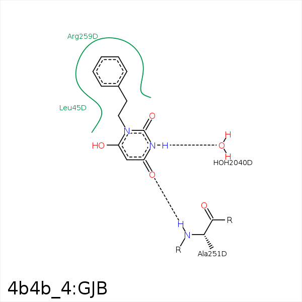


Represent the protein/ligand binding mode, centered on the ligand
Dashed lines represents hydrogen bonds and metal interactions
Green residue labels for amino acids with hydrophobic contacts (green lines) to the ligand
| Ligand | Protein | Interaction | |||
|---|---|---|---|---|---|
| Atom | Atom | Residue | Distance (Å) | Angle (°) | Type |
| CAQ | CD2 | LEU- 45 | 3.79 | 0 | Hydrophobic |
| OAN | N | ALA- 251 | 2.82 | 171.82 | H-Bond (Protein Donor) |
| CAF | CD | ARG- 259 | 3.38 | 0 | Hydrophobic |
| CAQ | CD | ARG- 259 | 4.14 | 0 | Hydrophobic |
| CAH | CB | ARG- 259 | 3.77 | 0 | Hydrophobic |
| NAB | O | HOH- 2040 | 2.94 | 169.56 | H-Bond (Ligand Donor) |
