sc-PDB
An Annotated Database of Druggable Binding Sites from the Protein DataBank
An Annotated Database of Druggable Binding Sites from the Protein DataBank
2.220 Å
X-ray
2013-02-20
| Name: | Muconolactone Delta-isomerase |
|---|---|
| ID: | Q8G9L0_RHOOP |
| AC: | Q8G9L0 |
| Organism: | Rhodococcus opacus |
| Reign: | Bacteria |
| TaxID: | 37919 |
| EC Number: | / |
| Chain Name: | Percentage of Residues within binding site |
|---|---|
| H | 14 % |
| J | 86 % |
| B-Factor: | 33.428 |
|---|---|
| Number of residues: | 26 |
| Including | |
| Standard Amino Acids: | 24 |
| Non Standard Amino Acids: | 1 |
| Water Molecules: | 1 |
| Cofactors: | |
| Metals: | CL |
| Ligandability | Volume (Å3) |
|---|---|
| 0.878 | 394.875 |
| % Hydrophobic | % Polar |
|---|---|
| 68.38 | 31.62 |
| According to VolSite | |
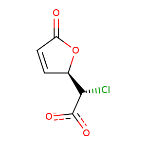
| HET Code: | K6H |
|---|---|
| Formula: | C6H4ClO4 |
| Molecular weight: | 175.547 g/mol |
| DrugBank ID: | - |
| Buried Surface Area: | 69.48 % |
| Polar Surface area: | 66.43 Å2 |
| Number of | |
|---|---|
| H-Bond Acceptors: | 4 |
| H-Bond Donors: | 0 |
| Rings: | 1 |
| Aromatic rings: | 0 |
| Anionic atoms: | 1 |
| Cationic atoms: | 0 |
| Rule of Five Violation: | 0 |
| Rotatable Bonds: | 2 |
| X | Y | Z |
|---|---|---|
| 41.6351 | 45.8944 | 37.7455 |
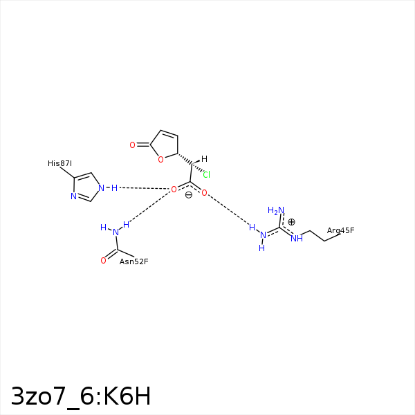


Represent the protein/ligand binding mode, centered on the ligand
Dashed lines represents hydrogen bonds and metal interactions
Green residue labels for amino acids with hydrophobic contacts (green lines) to the ligand
| Ligand | Protein | Interaction | |||
|---|---|---|---|---|---|
| Atom | Atom | Residue | Distance (Å) | Angle (°) | Type |
| CLAD | CB | MET- 7 | 4.36 | 0 | Hydrophobic |
| CLAD | CG2 | VAL- 9 | 3.56 | 0 | Hydrophobic |
| CAK | CB | ALA- 27 | 4.07 | 0 | Hydrophobic |
| OAC | CZ | ARG- 45 | 3.87 | 0 | Ionic (Protein Cationic) |
| OAC | NH1 | ARG- 45 | 3.02 | 152.08 | H-Bond (Protein Donor) |
| OAA | ND2 | ASN- 52 | 2.9 | 166.34 | H-Bond (Protein Donor) |
| CLAD | CD2 | PHE- 73 | 4.34 | 0 | Hydrophobic |
| CAK | CD2 | PHE- 73 | 3.42 | 0 | Hydrophobic |
| CLAD | CD2 | LEU- 77 | 3.88 | 0 | Hydrophobic |
| OAA | NE2 | HIS- 87 | 2.91 | 153.17 | H-Bond (Protein Donor) |
| OAC | NE2 | HIS- 87 | 3.48 | 135.77 | H-Bond (Protein Donor) |
