sc-PDB
An Annotated Database of Druggable Binding Sites from the Protein DataBank
An Annotated Database of Druggable Binding Sites from the Protein DataBank
2.650 Å
X-ray
2014-10-09
| Name: | Phosphatidylinositol 5-phosphate 4-kinase type-2 beta |
|---|---|
| ID: | PI42B_HUMAN |
| AC: | P78356 |
| Organism: | Homo sapiens |
| Reign: | Eukaryota |
| TaxID: | 9606 |
| EC Number: | 2.7.1.149 |
| Chain Name: | Percentage of Residues within binding site |
|---|---|
| A | 100 % |
| B-Factor: | 70.561 |
|---|---|
| Number of residues: | 21 |
| Including | |
| Standard Amino Acids: | 20 |
| Non Standard Amino Acids: | 0 |
| Water Molecules: | 1 |
| Cofactors: | |
| Metals: | |
| Ligandability | Volume (Å3) |
|---|---|
| 0.597 | 664.875 |
| % Hydrophobic | % Polar |
|---|---|
| 56.85 | 43.15 |
| According to VolSite | |
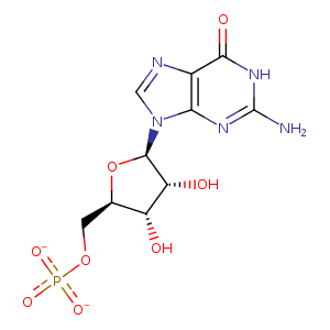
| HET Code: | 5GP |
|---|---|
| Formula: | C10H12N5O8P |
| Molecular weight: | 361.205 g/mol |
| DrugBank ID: | - |
| Buried Surface Area: | 42.33 % |
| Polar Surface area: | 217.22 Å2 |
| Number of | |
|---|---|
| H-Bond Acceptors: | 11 |
| H-Bond Donors: | 4 |
| Rings: | 3 |
| Aromatic rings: | 1 |
| Anionic atoms: | 2 |
| Cationic atoms: | 0 |
| Rule of Five Violation: | 1 |
| Rotatable Bonds: | 4 |
| X | Y | Z |
|---|---|---|
| 43.6945 | 60.7954 | -3.61213 |
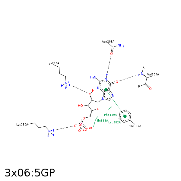


Represent the protein/ligand binding mode, centered on the ligand
Dashed lines represents hydrogen bonds and metal interactions
Green residue labels for amino acids with hydrophobic contacts (green lines) to the ligand
| Ligand | Protein | Interaction | |||
|---|---|---|---|---|---|
| Atom | Atom | Residue | Distance (Å) | Angle (°) | Type |
| C1' | CE2 | PHE- 139 | 4.16 | 0 | Hydrophobic |
| O3P | NZ | LYS- 150 | 3.19 | 145.76 | H-Bond (Protein Donor) |
| O3P | NZ | LYS- 150 | 3.19 | 0 | Ionic (Protein Cationic) |
| N1 | OD1 | ASN- 203 | 2.91 | 163.55 | H-Bond (Ligand Donor) |
| N2 | OD1 | ASN- 203 | 3.36 | 136.42 | H-Bond (Ligand Donor) |
| O6 | N | VAL- 204 | 3.08 | 147.92 | H-Bond (Protein Donor) |
| O2' | NZ | LYS- 214 | 2.94 | 153.94 | H-Bond (Protein Donor) |
| C2' | CD2 | LEU- 282 | 4.36 | 0 | Hydrophobic |
