sc-PDB
An Annotated Database of Druggable Binding Sites from the Protein DataBank
An Annotated Database of Druggable Binding Sites from the Protein DataBank
1.700 Å
X-ray
2011-11-25
| Name: | 4-hydroxy-3-methylbut-2-enyl diphosphate reductase |
|---|---|
| ID: | ISPH_ECOLI |
| AC: | P62623 |
| Organism: | Escherichia coli |
| Reign: | Bacteria |
| TaxID: | 83333 |
| EC Number: | / |
| Chain Name: | Percentage of Residues within binding site |
|---|---|
| B | 100 % |
| B-Factor: | 15.366 |
|---|---|
| Number of residues: | 31 |
| Including | |
| Standard Amino Acids: | 29 |
| Non Standard Amino Acids: | 1 |
| Water Molecules: | 1 |
| Cofactors: | |
| Metals: | |
| Ligandability | Volume (Å3) |
|---|---|
| 0.882 | 266.625 |
| % Hydrophobic | % Polar |
|---|---|
| 51.90 | 48.10 |
| According to VolSite | |
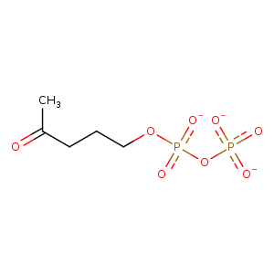
| HET Code: | 0CJ |
|---|---|
| Formula: | C5H9O8P2 |
| Molecular weight: | 259.068 g/mol |
| DrugBank ID: | - |
| Buried Surface Area: | 80.51 % |
| Polar Surface area: | 158.47 Å2 |
| Number of | |
|---|---|
| H-Bond Acceptors: | 8 |
| H-Bond Donors: | 0 |
| Rings: | 0 |
| Aromatic rings: | 0 |
| Anionic atoms: | 3 |
| Cationic atoms: | 0 |
| Rule of Five Violation: | 0 |
| Rotatable Bonds: | 7 |
| X | Y | Z |
|---|---|---|
| -38.4052 | -0.365067 | 15.8377 |
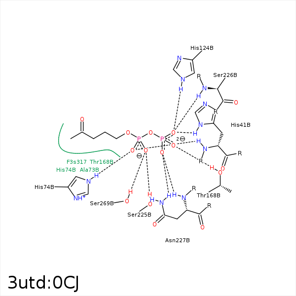


Represent the protein/ligand binding mode, centered on the ligand
Dashed lines represents hydrogen bonds and metal interactions
Green residue labels for amino acids with hydrophobic contacts (green lines) to the ligand
| Ligand | Protein | Interaction | |||
|---|---|---|---|---|---|
| Atom | Atom | Residue | Distance (Å) | Angle (°) | Type |
| C28 | CG2 | VAL- 15 | 3.32 | 0 | Hydrophobic |
| O15 | N | HIS- 41 | 2.76 | 176.29 | H-Bond (Protein Donor) |
| O19 | ND1 | HIS- 41 | 2.83 | 163.12 | H-Bond (Protein Donor) |
| C27 | CB | ALA- 73 | 4.26 | 0 | Hydrophobic |
| O15 | NE2 | HIS- 74 | 2.91 | 151.68 | H-Bond (Protein Donor) |
| O16 | NE2 | HIS- 74 | 3.3 | 123.98 | H-Bond (Protein Donor) |
| C22 | CB | CYS- 96 | 4.45 | 0 | Hydrophobic |
| C22 | CG2 | VAL- 99 | 3.42 | 0 | Hydrophobic |
| O19 | NE2 | HIS- 124 | 2.93 | 167.14 | H-Bond (Protein Donor) |
| C30 | CG2 | THR- 168 | 3.56 | 0 | Hydrophobic |
| O18 | OG1 | THR- 168 | 2.84 | 158.92 | H-Bond (Protein Donor) |
| O14 | OG | SER- 225 | 2.64 | 145.14 | H-Bond (Protein Donor) |
| O19 | N | SER- 226 | 2.63 | 156.7 | H-Bond (Protein Donor) |
| O20 | N | ASN- 227 | 2.72 | 167.26 | H-Bond (Protein Donor) |
| O20 | ND2 | ASN- 227 | 2.83 | 154.78 | H-Bond (Protein Donor) |
| C28 | CB | SER- 269 | 4.3 | 0 | Hydrophobic |
| O14 | OG | SER- 269 | 2.72 | 156.93 | H-Bond (Protein Donor) |
