sc-PDB
An Annotated Database of Druggable Binding Sites from the Protein DataBank
An Annotated Database of Druggable Binding Sites from the Protein DataBank
2.000 Å
X-ray
2011-11-18
| Name: | UDP-N-acetylglucosamine 1-carboxyvinyltransferase |
|---|---|
| ID: | MURA_ENTCC |
| AC: | P33038 |
| Organism: | Enterobacter cloacae subsp. cloacae |
| Reign: | Bacteria |
| TaxID: | 716541 |
| EC Number: | / |
| Chain Name: | Percentage of Residues within binding site |
|---|---|
| A | 100 % |
| B-Factor: | 19.477 |
|---|---|
| Number of residues: | 43 |
| Including | |
| Standard Amino Acids: | 41 |
| Non Standard Amino Acids: | 0 |
| Water Molecules: | 2 |
| Cofactors: | |
| Metals: | |
| Ligandability | Volume (Å3) |
|---|---|
| 0.791 | 810.000 |
| % Hydrophobic | % Polar |
|---|---|
| 48.33 | 51.67 |
| According to VolSite | |
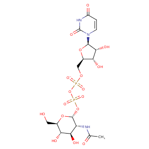
| HET Code: | UD1 |
|---|---|
| Formula: | C17H25N3O17P2 |
| Molecular weight: | 605.338 g/mol |
| DrugBank ID: | DB03397 |
| Buried Surface Area: | 60.52 % |
| Polar Surface area: | 325.69 Å2 |
| Number of | |
|---|---|
| H-Bond Acceptors: | 17 |
| H-Bond Donors: | 7 |
| Rings: | 3 |
| Aromatic rings: | 0 |
| Anionic atoms: | 2 |
| Cationic atoms: | 0 |
| Rule of Five Violation: | 3 |
| Rotatable Bonds: | 10 |
| X | Y | Z |
|---|---|---|
| 83.6946 | 3.38441 | 126.39 |


Represent the protein/ligand binding mode, centered on the ligand
Dashed lines represents hydrogen bonds and metal interactions
Green residue labels for amino acids with hydrophobic contacts (green lines) to the ligand
| Ligand | Protein | Interaction | |||
|---|---|---|---|---|---|
| Atom | Atom | Residue | Distance (Å) | Angle (°) | Type |
| O3' | ND2 | ASN- 23 | 3.43 | 125.69 | H-Bond (Protein Donor) |
| C8' | CD1 | LEU- 26 | 4.07 | 0 | Hydrophobic |
| C8' | CB | ALA- 92 | 3.97 | 0 | Hydrophobic |
| C8' | CH2 | TRP- 95 | 3.5 | 0 | Hydrophobic |
| O2B | NH2 | ARG- 120 | 3.01 | 160.04 | H-Bond (Protein Donor) |
| O2B | CZ | ARG- 120 | 3.97 | 0 | Ionic (Protein Cationic) |
| N3 | OD1 | ASP- 123 | 2.66 | 161.48 | H-Bond (Ligand Donor) |
| O4 | N | LEU- 124 | 2.71 | 142.64 | H-Bond (Protein Donor) |
| O2 | NZ | LYS- 160 | 3.21 | 157.25 | H-Bond (Protein Donor) |
| C1B | CD | LYS- 160 | 3.97 | 0 | Hydrophobic |
| O2A | OG | SER- 162 | 2.61 | 172.9 | H-Bond (Protein Donor) |
| C1' | CG1 | VAL- 163 | 4.02 | 0 | Hydrophobic |
| O1A | N | VAL- 163 | 2.76 | 159.84 | H-Bond (Protein Donor) |
| O2A | N | GLY- 164 | 3.33 | 149.09 | H-Bond (Protein Donor) |
| O1B | N | GLY- 164 | 2.89 | 122.51 | H-Bond (Protein Donor) |
| C6' | CG2 | THR- 304 | 3.64 | 0 | Hydrophobic |
| O3' | OD2 | ASP- 305 | 3.07 | 164.35 | H-Bond (Ligand Donor) |
| O4' | OD2 | ASP- 305 | 3.23 | 137.46 | H-Bond (Ligand Donor) |
| O4' | OD1 | ASP- 305 | 2.65 | 158.6 | H-Bond (Ligand Donor) |
| C6' | CG2 | ILE- 327 | 4.26 | 0 | Hydrophobic |
| C5B | CG2 | ILE- 327 | 3.92 | 0 | Hydrophobic |
| O3B | O | ILE- 327 | 2.92 | 161.44 | H-Bond (Ligand Donor) |
| C5' | CE2 | PHE- 328 | 3.9 | 0 | Hydrophobic |
| C3B | CE1 | PHE- 328 | 3.61 | 0 | Hydrophobic |
