sc-PDB
An Annotated Database of Druggable Binding Sites from the Protein DataBank
An Annotated Database of Druggable Binding Sites from the Protein DataBank
1.990 Å
X-ray
2011-10-24
| Name: | Formyl-CoA:oxalate CoA-transferase |
|---|---|
| ID: | A9X6P7_ACEAC |
| AC: | A9X6P7 |
| Organism: | Acetobacter aceti |
| Reign: | Bacteria |
| TaxID: | 435 |
| EC Number: | / |
| Chain Name: | Percentage of Residues within binding site |
|---|---|
| D | 100 % |
| B-Factor: | 25.173 |
|---|---|
| Number of residues: | 41 |
| Including | |
| Standard Amino Acids: | 41 |
| Non Standard Amino Acids: | 0 |
| Water Molecules: | 0 |
| Cofactors: | |
| Metals: | |
| Ligandability | Volume (Å3) |
|---|---|
| 0.431 | 722.250 |
| % Hydrophobic | % Polar |
|---|---|
| 43.46 | 56.54 |
| According to VolSite | |
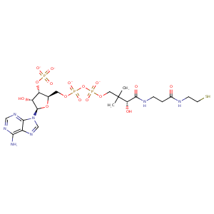
| HET Code: | COA |
|---|---|
| Formula: | C21H32N7O16P3S |
| Molecular weight: | 763.502 g/mol |
| DrugBank ID: | DB01992 |
| Buried Surface Area: | 63.46 % |
| Polar Surface area: | 426.11 Å2 |
| Number of | |
|---|---|
| H-Bond Acceptors: | 21 |
| H-Bond Donors: | 6 |
| Rings: | 3 |
| Aromatic rings: | 2 |
| Anionic atoms: | 4 |
| Cationic atoms: | 0 |
| Rule of Five Violation: | 3 |
| Rotatable Bonds: | 18 |
| X | Y | Z |
|---|---|---|
| -33.5741 | -7.03398 | 129.386 |
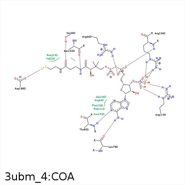


Represent the protein/ligand binding mode, centered on the ligand
Dashed lines represents hydrogen bonds and metal interactions
Green residue labels for amino acids with hydrophobic contacts (green lines) to the ligand
| Ligand | Protein | Interaction | |||
|---|---|---|---|---|---|
| Atom | Atom | Residue | Distance (Å) | Angle (°) | Type |
| CEP | CG2 | VAL- 22 | 4.07 | 0 | Hydrophobic |
| C2P | CB | VAL- 22 | 3.67 | 0 | Hydrophobic |
| O5P | OG | SER- 24 | 3.06 | 140.88 | H-Bond (Protein Donor) |
| O1A | CZ | ARG- 44 | 3.95 | 0 | Ionic (Protein Cationic) |
| O1A | NH2 | ARG- 44 | 3.19 | 160.85 | H-Bond (Protein Donor) |
| N6A | O | LEU- 78 | 2.77 | 166.18 | H-Bond (Ligand Donor) |
| N1A | N | THR- 80 | 3.17 | 153.03 | H-Bond (Protein Donor) |
| O3B | NZ | LYS- 81 | 3.48 | 133.78 | H-Bond (Protein Donor) |
| O7A | NZ | LYS- 81 | 3.9 | 0 | Ionic (Protein Cationic) |
| O8A | NZ | LYS- 81 | 3.54 | 0 | Ionic (Protein Cationic) |
| N8P | O | ASN- 102 | 2.64 | 145.25 | H-Bond (Ligand Donor) |
| O5P | ND2 | ASN- 102 | 3.45 | 140.35 | H-Bond (Protein Donor) |
| C2B | CG | PHE- 103 | 4.43 | 0 | Hydrophobic |
| O8A | NH2 | ARG- 104 | 2.59 | 143.61 | H-Bond (Protein Donor) |
| O9A | NH2 | ARG- 104 | 2.96 | 122.42 | H-Bond (Protein Donor) |
| O2A | N | ARG- 104 | 2.88 | 147.16 | H-Bond (Protein Donor) |
| O8A | CZ | ARG- 104 | 3.63 | 0 | Ionic (Protein Cationic) |
| O9A | CZ | ARG- 104 | 3.06 | 0 | Ionic (Protein Cationic) |
| CAP | CB | ARG- 104 | 4.11 | 0 | Hydrophobic |
| C3B | CB | ALA- 107 | 3.85 | 0 | Hydrophobic |
| O7A | CZ | ARG- 110 | 3.41 | 0 | Ionic (Protein Cationic) |
| O9A | CZ | ARG- 110 | 3.57 | 0 | Ionic (Protein Cationic) |
| O7A | NH2 | ARG- 110 | 3.43 | 123.97 | H-Bond (Protein Donor) |
| O7A | NE | ARG- 110 | 2.56 | 170.36 | H-Bond (Protein Donor) |
| O9A | NH2 | ARG- 110 | 2.7 | 164.95 | H-Bond (Protein Donor) |
| C2B | CE | MET- 111 | 4.16 | 0 | Hydrophobic |
| C6P | CG2 | VAL- 130 | 4 | 0 | Hydrophobic |
| C6P | CB | ALA- 144 | 3.87 | 0 | Hydrophobic |
| CDP | CE2 | TYR- 145 | 4.12 | 0 | Hydrophobic |
| C6P | SD | MET- 219 | 3.31 | 0 | Hydrophobic |
| S1P | CE | MET- 219 | 3.25 | 0 | Hydrophobic |
