sc-PDB
An Annotated Database of Druggable Binding Sites from the Protein DataBank
An Annotated Database of Druggable Binding Sites from the Protein DataBank
2.400 Å
X-ray
2011-07-25
| Name: | Cytochrome P450 2A6 |
|---|---|
| ID: | CP2A6_HUMAN |
| AC: | P11509 |
| Organism: | Homo sapiens |
| Reign: | Eukaryota |
| TaxID: | 9606 |
| EC Number: | 1.14.13 |
| Chain Name: | Percentage of Residues within binding site |
|---|---|
| B | 100 % |
| B-Factor: | 21.411 |
|---|---|
| Number of residues: | 21 |
| Including | |
| Standard Amino Acids: | 20 |
| Non Standard Amino Acids: | 1 |
| Water Molecules: | 0 |
| Cofactors: | |
| Metals: | |
| Ligandability | Volume (Å3) |
|---|---|
| 1.592 | 1032.750 |
| % Hydrophobic | % Polar |
|---|---|
| 61.11 | 38.89 |
| According to VolSite | |
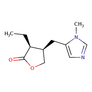
| HET Code: | 9PL |
|---|---|
| Formula: | C11H16N2O2 |
| Molecular weight: | 208.257 g/mol |
| DrugBank ID: | DB01085 |
| Buried Surface Area: | 83.72 % |
| Polar Surface area: | 44.12 Å2 |
| Number of | |
|---|---|
| H-Bond Acceptors: | 3 |
| H-Bond Donors: | 0 |
| Rings: | 2 |
| Aromatic rings: | 1 |
| Anionic atoms: | 0 |
| Cationic atoms: | 0 |
| Rule of Five Violation: | 0 |
| Rotatable Bonds: | 3 |
| X | Y | Z |
|---|---|---|
| -22.2707 | 31.9061 | -7.891 |
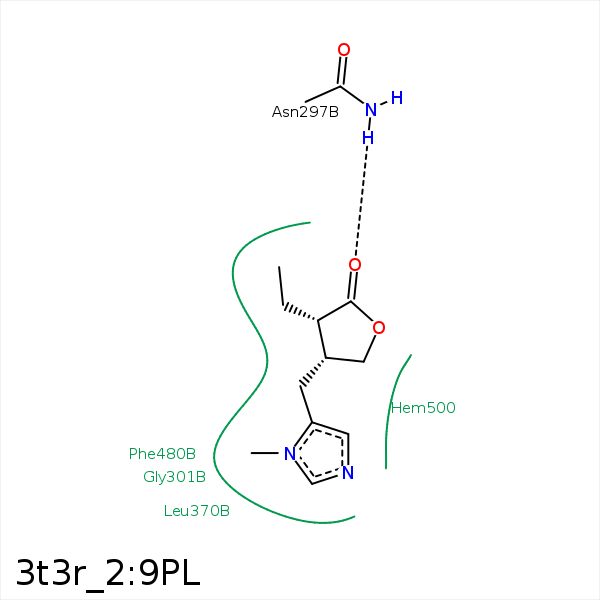


Represent the protein/ligand binding mode, centered on the ligand
Dashed lines represents hydrogen bonds and metal interactions
Green residue labels for amino acids with hydrophobic contacts (green lines) to the ligand
| Ligand | Protein | Interaction | |||
|---|---|---|---|---|---|
| Atom | Atom | Residue | Distance (Å) | Angle (°) | Type |
| C12 | CD2 | PHE- 107 | 4.1 | 0 | Hydrophobic |
| C13 | CG | PHE- 107 | 3.82 | 0 | Hydrophobic |
| C14 | CD1 | PHE- 107 | 3.36 | 0 | Hydrophobic |
| C9 | CG2 | VAL- 117 | 4.01 | 0 | Hydrophobic |
| C13 | CG1 | VAL- 117 | 4.37 | 0 | Hydrophobic |
| C13 | CE1 | PHE- 118 | 3.83 | 0 | Hydrophobic |
| C14 | CZ | PHE- 118 | 4.07 | 0 | Hydrophobic |
| C6 | CZ | PHE- 209 | 3.31 | 0 | Hydrophobic |
| C7 | CZ | PHE- 209 | 4.47 | 0 | Hydrophobic |
| O15 | ND2 | ASN- 297 | 3.13 | 160.18 | H-Bond (Protein Donor) |
| C6 | CG2 | ILE- 300 | 4.46 | 0 | Hydrophobic |
| C12 | CG2 | ILE- 300 | 4.07 | 0 | Hydrophobic |
| C6 | CB | GLU- 304 | 4.25 | 0 | Hydrophobic |
| C6 | CG2 | THR- 305 | 3.5 | 0 | Hydrophobic |
| C7 | CD1 | LEU- 370 | 4.24 | 0 | Hydrophobic |
| C7 | CZ | PHE- 480 | 4.33 | 0 | Hydrophobic |
| C14 | CZ | PHE- 480 | 3.61 | 0 | Hydrophobic |
