sc-PDB
An Annotated Database of Druggable Binding Sites from the Protein DataBank
An Annotated Database of Druggable Binding Sites from the Protein DataBank
2.650 Å
X-ray
2011-07-25
| Name: | Glycogen phosphorylase, muscle form |
|---|---|
| ID: | PYGM_RABIT |
| AC: | P00489 |
| Organism: | Oryctolagus cuniculus |
| Reign: | Eukaryota |
| TaxID: | 9986 |
| EC Number: | 2.4.1.1 |
| Chain Name: | Percentage of Residues within binding site |
|---|---|
| A | 100 % |
| B-Factor: | 17.215 |
|---|---|
| Number of residues: | 37 |
| Including | |
| Standard Amino Acids: | 34 |
| Non Standard Amino Acids: | 1 |
| Water Molecules: | 2 |
| Cofactors: | |
| Metals: | |
| Ligandability | Volume (Å3) |
|---|---|
| 0.738 | 533.250 |
| % Hydrophobic | % Polar |
|---|---|
| 48.73 | 51.27 |
| According to VolSite | |
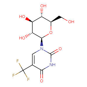
| HET Code: | GPW |
|---|---|
| Formula: | C11H13F3N2O7 |
| Molecular weight: | 342.225 g/mol |
| DrugBank ID: | - |
| Buried Surface Area: | 77.52 % |
| Polar Surface area: | 139.56 Å2 |
| Number of | |
|---|---|
| H-Bond Acceptors: | 7 |
| H-Bond Donors: | 5 |
| Rings: | 2 |
| Aromatic rings: | 0 |
| Anionic atoms: | 0 |
| Cationic atoms: | 0 |
| Rule of Five Violation: | 0 |
| Rotatable Bonds: | 3 |
| X | Y | Z |
|---|---|---|
| 34.4359 | 22.9822 | 28.5337 |
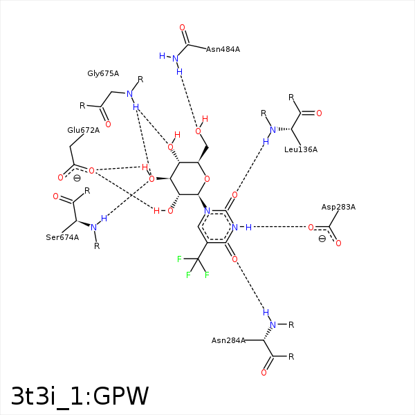


Represent the protein/ligand binding mode, centered on the ligand
Dashed lines represents hydrogen bonds and metal interactions
Green residue labels for amino acids with hydrophobic contacts (green lines) to the ligand
| Ligand | Protein | Interaction | |||
|---|---|---|---|---|---|
| Atom | Atom | Residue | Distance (Å) | Angle (°) | Type |
| O2 | N | LEU- 136 | 3.19 | 147.92 | H-Bond (Protein Donor) |
| C1' | CB | LEU- 136 | 4.1 | 0 | Hydrophobic |
| C6' | CB | LEU- 136 | 4.47 | 0 | Hydrophobic |
| F2 | CD1 | LEU- 136 | 3.76 | 0 | Hydrophobic |
| C6' | CB | LEU- 139 | 4.18 | 0 | Hydrophobic |
| N3 | OD1 | ASP- 283 | 2.73 | 162.68 | H-Bond (Ligand Donor) |
| F1 | CB | ASN- 284 | 4.38 | 0 | Hydrophobic |
| O4 | N | ASN- 284 | 2.94 | 158.45 | H-Bond (Protein Donor) |
| O2' | ND2 | ASN- 284 | 3.47 | 158.05 | H-Bond (Protein Donor) |
| F2 | CB | ASP- 339 | 4.34 | 0 | Hydrophobic |
| C7 | CB | HIS- 377 | 4.22 | 0 | Hydrophobic |
| F1 | CG2 | THR- 378 | 3.6 | 0 | Hydrophobic |
| F3 | CB | THR- 378 | 3.49 | 0 | Hydrophobic |
| F1 | CB | ALA- 383 | 3.62 | 0 | Hydrophobic |
| O4' | ND2 | ASN- 484 | 3.39 | 125.91 | H-Bond (Protein Donor) |
| O2' | OE1 | GLU- 672 | 3.12 | 157.6 | H-Bond (Ligand Donor) |
| O3' | OE1 | GLU- 672 | 2.68 | 156.87 | H-Bond (Ligand Donor) |
| O3' | N | SER- 674 | 2.93 | 167.89 | H-Bond (Protein Donor) |
| C4' | CB | SER- 674 | 4.31 | 0 | Hydrophobic |
| O3' | N | GLY- 675 | 3.15 | 145.68 | H-Bond (Protein Donor) |
| O4' | N | GLY- 675 | 2.84 | 129.83 | H-Bond (Protein Donor) |
