sc-PDB
An Annotated Database of Druggable Binding Sites from the Protein DataBank
An Annotated Database of Druggable Binding Sites from the Protein DataBank
2.800 Å
X-ray
2011-01-26
| Name: | Guanine nucleotide-binding protein G(i) subunit alpha-1 |
|---|---|
| ID: | GNAI1_HUMAN |
| AC: | P63096 |
| Organism: | Homo sapiens |
| Reign: | Eukaryota |
| TaxID: | 9606 |
| EC Number: | / |
| Chain Name: | Percentage of Residues within binding site |
|---|---|
| B | 97 % |
| D | 3 % |
| B-Factor: | 58.294 |
|---|---|
| Number of residues: | 35 |
| Including | |
| Standard Amino Acids: | 35 |
| Non Standard Amino Acids: | 0 |
| Water Molecules: | 0 |
| Cofactors: | |
| Metals: | |
| Ligandability | Volume (Å3) |
|---|---|
| 0.707 | 513.000 |
| % Hydrophobic | % Polar |
|---|---|
| 49.34 | 50.66 |
| According to VolSite | |
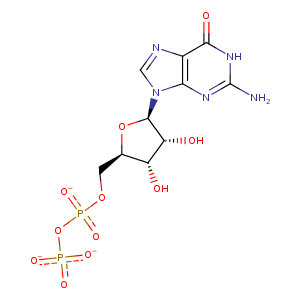
| HET Code: | GDP |
|---|---|
| Formula: | C10H12N5O11P2 |
| Molecular weight: | 440.177 g/mol |
| DrugBank ID: | DB04315 |
| Buried Surface Area: | 80.03 % |
| Polar Surface area: | 276.39 Å2 |
| Number of | |
|---|---|
| H-Bond Acceptors: | 14 |
| H-Bond Donors: | 4 |
| Rings: | 3 |
| Aromatic rings: | 1 |
| Anionic atoms: | 3 |
| Cationic atoms: | 0 |
| Rule of Five Violation: | 1 |
| Rotatable Bonds: | 6 |
| X | Y | Z |
|---|---|---|
| -22.4284 | 55.7284 | 14.4364 |
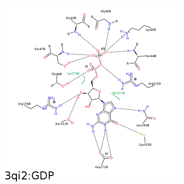


Represent the protein/ligand binding mode, centered on the ligand
Dashed lines represents hydrogen bonds and metal interactions
Green residue labels for amino acids with hydrophobic contacts (green lines) to the ligand
| Ligand | Protein | Interaction | |||
|---|---|---|---|---|---|
| Atom | Atom | Residue | Distance (Å) | Angle (°) | Type |
| O2B | N | GLU- 43 | 3.16 | 173.93 | H-Bond (Protein Donor) |
| C5' | CB | GLU- 43 | 4.42 | 0 | Hydrophobic |
| O3B | N | SER- 44 | 2.94 | 130.23 | H-Bond (Protein Donor) |
| O3B | N | GLY- 45 | 3.13 | 171.74 | H-Bond (Protein Donor) |
| O1B | NZ | LYS- 46 | 3.85 | 0 | Ionic (Protein Cationic) |
| O2B | NZ | LYS- 46 | 3.31 | 0 | Ionic (Protein Cationic) |
| O3B | NZ | LYS- 46 | 2.99 | 0 | Ionic (Protein Cationic) |
| O3B | N | LYS- 46 | 3.28 | 134.02 | H-Bond (Protein Donor) |
| O3B | NZ | LYS- 46 | 2.99 | 158.78 | H-Bond (Protein Donor) |
| O1B | OG | SER- 47 | 2.72 | 153.88 | H-Bond (Protein Donor) |
| O1B | N | SER- 47 | 2.86 | 164.58 | H-Bond (Protein Donor) |
| O1A | N | THR- 48 | 3.41 | 121.15 | H-Bond (Protein Donor) |
| O1A | OG1 | THR- 48 | 2.74 | 139.27 | H-Bond (Protein Donor) |
| C4' | CB | ASP- 150 | 4.49 | 0 | Hydrophobic |
| C1' | CB | ASP- 150 | 3.79 | 0 | Hydrophobic |
| O3' | OG | SER- 151 | 2.7 | 149.15 | H-Bond (Ligand Donor) |
| C2' | CD | ARG- 176 | 4.35 | 0 | Hydrophobic |
| O3' | NH1 | ARG- 178 | 3.14 | 133.46 | H-Bond (Protein Donor) |
| N7 | ND2 | ASN- 269 | 3 | 131.4 | H-Bond (Protein Donor) |
| O4' | NZ | LYS- 270 | 3.46 | 123.12 | H-Bond (Protein Donor) |
| O6 | N | LYS- 270 | 3.42 | 122.47 | H-Bond (Protein Donor) |
| N1 | OD1 | ASP- 272 | 2.73 | 143.19 | H-Bond (Ligand Donor) |
| N1 | OD2 | ASP- 272 | 3.15 | 136.45 | H-Bond (Ligand Donor) |
| N2 | OD1 | ASP- 272 | 3.5 | 121.64 | H-Bond (Ligand Donor) |
| N2 | OD2 | ASP- 272 | 2.78 | 153.77 | H-Bond (Ligand Donor) |
| C2' | CG2 | THR- 327 | 4.02 | 0 | Hydrophobic |
| O3A | NH1 | ARG- 516 | 3.19 | 167.76 | H-Bond (Protein Donor) |
