sc-PDB
An Annotated Database of Druggable Binding Sites from the Protein DataBank
An Annotated Database of Druggable Binding Sites from the Protein DataBank
1.100 Å
X-ray
2011-01-25
| Name: | Histidine triad nucleotide-binding protein 1 |
|---|---|
| ID: | HINT1_RABIT |
| AC: | P80912 |
| Organism: | Oryctolagus cuniculus |
| Reign: | Eukaryota |
| TaxID: | 9986 |
| EC Number: | 3 |
| Chain Name: | Percentage of Residues within binding site |
|---|---|
| A | 100 % |
| B-Factor: | 14.517 |
|---|---|
| Number of residues: | 24 |
| Including | |
| Standard Amino Acids: | 23 |
| Non Standard Amino Acids: | 0 |
| Water Molecules: | 1 |
| Cofactors: | |
| Metals: | |
| Ligandability | Volume (Å3) |
|---|---|
| 0.629 | 327.375 |
| % Hydrophobic | % Polar |
|---|---|
| 61.86 | 38.14 |
| According to VolSite | |
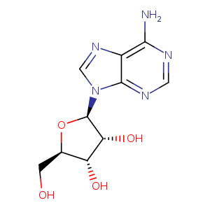
| HET Code: | ADN |
|---|---|
| Formula: | C10H13N5O4 |
| Molecular weight: | 267.241 g/mol |
| DrugBank ID: | DB00640 |
| Buried Surface Area: | 55.25 % |
| Polar Surface area: | 139.54 Å2 |
| Number of | |
|---|---|
| H-Bond Acceptors: | 8 |
| H-Bond Donors: | 4 |
| Rings: | 3 |
| Aromatic rings: | 2 |
| Anionic atoms: | 0 |
| Cationic atoms: | 0 |
| Rule of Five Violation: | 0 |
| Rotatable Bonds: | 2 |
| X | Y | Z |
|---|---|---|
| 8.24368 | 10.6281 | 55.3425 |
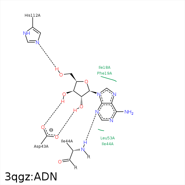


Represent the protein/ligand binding mode, centered on the ligand
Dashed lines represents hydrogen bonds and metal interactions
Green residue labels for amino acids with hydrophobic contacts (green lines) to the ligand
| Ligand | Protein | Interaction | |||
|---|---|---|---|---|---|
| Atom | Atom | Residue | Distance (Å) | Angle (°) | Type |
| C1' | CZ | PHE- 19 | 4.37 | 0 | Hydrophobic |
| C5' | CZ | PHE- 19 | 3.98 | 0 | Hydrophobic |
| O3' | OD2 | ASP- 43 | 2.58 | 163.5 | H-Bond (Ligand Donor) |
| O2' | OD1 | ASP- 43 | 2.83 | 169.86 | H-Bond (Ligand Donor) |
| N3 | N | ILE- 44 | 3.21 | 153.76 | H-Bond (Protein Donor) |
| C2' | CG2 | ILE- 44 | 4.18 | 0 | Hydrophobic |
| C4' | CD1 | LEU- 53 | 4 | 0 | Hydrophobic |
| C1' | CD1 | LEU- 53 | 4.07 | 0 | Hydrophobic |
| C5' | CG2 | VAL- 108 | 4.21 | 0 | Hydrophobic |
| O5' | NE2 | HIS- 112 | 2.52 | 136.36 | H-Bond (Ligand Donor) |
