sc-PDB
An Annotated Database of Druggable Binding Sites from the Protein DataBank
An Annotated Database of Druggable Binding Sites from the Protein DataBank
2.200 Å
X-ray
2011-01-17
| Name: | 3-phosphoinositide-dependent protein kinase 1 |
|---|---|
| ID: | PDPK1_HUMAN |
| AC: | O15530 |
| Organism: | Homo sapiens |
| Reign: | Eukaryota |
| TaxID: | 9606 |
| EC Number: | 2.7.11.1 |
| Chain Name: | Percentage of Residues within binding site |
|---|---|
| A | 100 % |
| B-Factor: | 24.394 |
|---|---|
| Number of residues: | 39 |
| Including | |
| Standard Amino Acids: | 35 |
| Non Standard Amino Acids: | 0 |
| Water Molecules: | 4 |
| Cofactors: | |
| Metals: | |
| Ligandability | Volume (Å3) |
|---|---|
| 0.673 | 492.750 |
| % Hydrophobic | % Polar |
|---|---|
| 51.37 | 48.63 |
| According to VolSite | |

| HET Code: | 3Q3 |
|---|---|
| Formula: | C22H22N8O2 |
| Molecular weight: | 430.462 g/mol |
| DrugBank ID: | - |
| Buried Surface Area: | 61.04 % |
| Polar Surface area: | 148.07 Å2 |
| Number of | |
|---|---|
| H-Bond Acceptors: | 8 |
| H-Bond Donors: | 4 |
| Rings: | 5 |
| Aromatic rings: | 4 |
| Anionic atoms: | 0 |
| Cationic atoms: | 0 |
| Rule of Five Violation: | 0 |
| Rotatable Bonds: | 4 |
| X | Y | Z |
|---|---|---|
| -45.4654 | 19.3403 | 11.3879 |
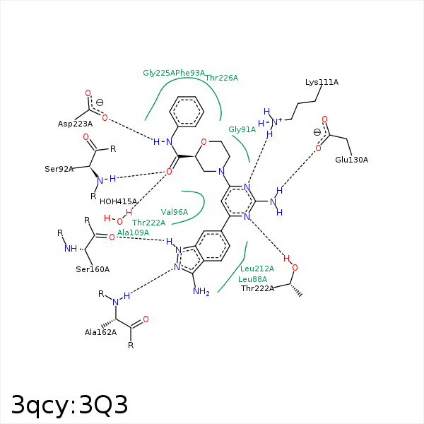


Represent the protein/ligand binding mode, centered on the ligand
Dashed lines represents hydrogen bonds and metal interactions
Green residue labels for amino acids with hydrophobic contacts (green lines) to the ligand
| Ligand | Protein | Interaction | |||
|---|---|---|---|---|---|
| Atom | Atom | Residue | Distance (Å) | Angle (°) | Type |
| C4 | CB | LEU- 88 | 4.19 | 0 | Hydrophobic |
| O31 | N | SER- 92 | 3.12 | 158.69 | H-Bond (Protein Donor) |
| C7 | CB | SER- 92 | 4.16 | 0 | Hydrophobic |
| C9 | CG2 | VAL- 96 | 3.72 | 0 | Hydrophobic |
| C4 | CG1 | VAL- 96 | 4.42 | 0 | Hydrophobic |
| C11 | CG2 | VAL- 96 | 3.99 | 0 | Hydrophobic |
| C8 | CG1 | VAL- 96 | 4.2 | 0 | Hydrophobic |
| C12 | CB | ALA- 109 | 3.88 | 0 | Hydrophobic |
| N24 | NZ | LYS- 111 | 3 | 171.71 | H-Bond (Protein Donor) |
| C2 | CB | TYR- 126 | 4.17 | 0 | Hydrophobic |
| N29 | OE2 | GLU- 130 | 2.86 | 163.86 | H-Bond (Ligand Donor) |
| C8 | CD1 | LEU- 159 | 4.12 | 0 | Hydrophobic |
| N26 | O | SER- 160 | 2.93 | 135.53 | H-Bond (Ligand Donor) |
| N25 | N | ALA- 162 | 2.95 | 176.42 | H-Bond (Protein Donor) |
| N28 | O | ALA- 162 | 3.24 | 128.27 | H-Bond (Ligand Donor) |
| C10 | CD1 | LEU- 212 | 3.36 | 0 | Hydrophobic |
| C12 | CD1 | LEU- 212 | 3.36 | 0 | Hydrophobic |
| C9 | CG2 | THR- 222 | 4.39 | 0 | Hydrophobic |
| C11 | CG2 | THR- 222 | 4.13 | 0 | Hydrophobic |
| N23 | OG1 | THR- 222 | 2.94 | 151.75 | H-Bond (Protein Donor) |
| N30 | OD1 | ASP- 223 | 2.75 | 147.09 | H-Bond (Ligand Donor) |
| C2 | CG2 | THR- 226 | 3.35 | 0 | Hydrophobic |
| O31 | O | HOH- 415 | 2.71 | 165.4 | H-Bond (Protein Donor) |
