sc-PDB
An Annotated Database of Druggable Binding Sites from the Protein DataBank
An Annotated Database of Druggable Binding Sites from the Protein DataBank
2.000 Å
X-ray
2010-11-03
| Name: | Purine nucleoside phosphorylase |
|---|---|
| ID: | Q8I3X4_PLAF7 |
| AC: | Q8I3X4 |
| Organism: | Plasmodium falciparum |
| Reign: | Eukaryota |
| TaxID: | 36329 |
| EC Number: | / |
| Chain Name: | Percentage of Residues within binding site |
|---|---|
| A | 86 % |
| B | 14 % |
| B-Factor: | 30.735 |
|---|---|
| Number of residues: | 37 |
| Including | |
| Standard Amino Acids: | 36 |
| Non Standard Amino Acids: | 0 |
| Water Molecules: | 1 |
| Cofactors: | |
| Metals: | |
| Ligandability | Volume (Å3) |
|---|---|
| 0.409 | 675.000 |
| % Hydrophobic | % Polar |
|---|---|
| 39.00 | 61.00 |
| According to VolSite | |
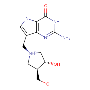
| HET Code: | IM5 |
|---|---|
| Formula: | C12H18N5O3 |
| Molecular weight: | 280.303 g/mol |
| DrugBank ID: | - |
| Buried Surface Area: | 74.28 % |
| Polar Surface area: | 128.17 Å2 |
| Number of | |
|---|---|
| H-Bond Acceptors: | 5 |
| H-Bond Donors: | 6 |
| Rings: | 3 |
| Aromatic rings: | 1 |
| Anionic atoms: | 0 |
| Cationic atoms: | 1 |
| Rule of Five Violation: | 1 |
| Rotatable Bonds: | 3 |
| X | Y | Z |
|---|---|---|
| -16.7917 | 9.76185 | -18.9069 |
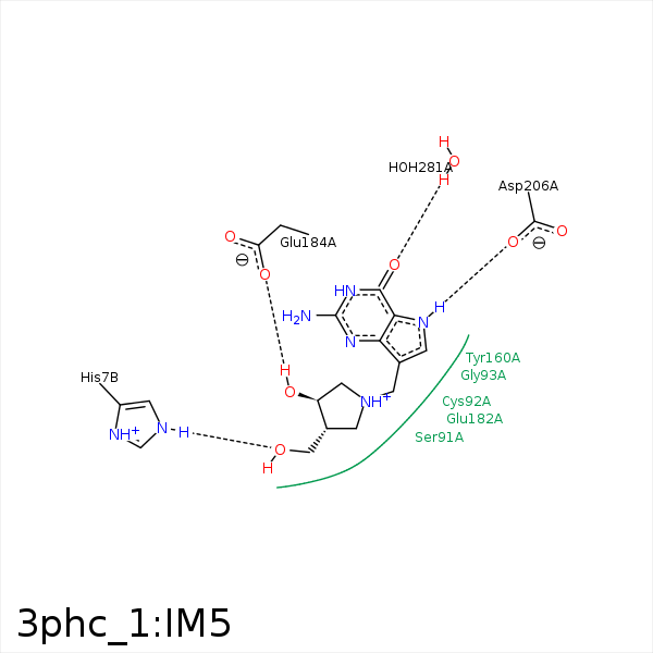


Represent the protein/ligand binding mode, centered on the ligand
Dashed lines represents hydrogen bonds and metal interactions
Green residue labels for amino acids with hydrophobic contacts (green lines) to the ligand
| Ligand | Protein | Interaction | |||
|---|---|---|---|---|---|
| Atom | Atom | Residue | Distance (Å) | Angle (°) | Type |
| O5' | NE2 | HIS- 7 | 2.54 | 163.49 | H-Bond (Protein Donor) |
| C3' | CG1 | VAL- 66 | 3.78 | 0 | Hydrophobic |
| C4' | CG2 | VAL- 66 | 4.31 | 0 | Hydrophobic |
| C5' | CG1 | VAL- 66 | 4.25 | 0 | Hydrophobic |
| C5' | CE1 | TYR- 160 | 3.9 | 0 | Hydrophobic |
| C3' | SD | MET- 183 | 3.68 | 0 | Hydrophobic |
| C5' | SD | MET- 183 | 4.2 | 0 | Hydrophobic |
| O3' | OE2 | GLU- 184 | 2.66 | 148.99 | H-Bond (Ligand Donor) |
| N7 | OD1 | ASP- 206 | 2.83 | 151.32 | H-Bond (Ligand Donor) |
| O6 | O | HOH- 281 | 2.76 | 165.1 | H-Bond (Protein Donor) |
