sc-PDB
An Annotated Database of Druggable Binding Sites from the Protein DataBank
An Annotated Database of Druggable Binding Sites from the Protein DataBank
2.410 Å
X-ray
2010-09-21
| Name: | Ribosyldihydronicotinamide dehydrogenase [quinone] |
|---|---|
| ID: | NQO2_HUMAN |
| AC: | P16083 |
| Organism: | Homo sapiens |
| Reign: | Eukaryota |
| TaxID: | 9606 |
| EC Number: | / |
| Chain Name: | Percentage of Residues within binding site |
|---|---|
| A | 57 % |
| B | 43 % |
| B-Factor: | 31.108 |
|---|---|
| Number of residues: | 31 |
| Including | |
| Standard Amino Acids: | 29 |
| Non Standard Amino Acids: | 1 |
| Water Molecules: | 1 |
| Cofactors: | FAD |
| Metals: | |
| Ligandability | Volume (Å3) |
|---|---|
| 0.847 | 577.125 |
| % Hydrophobic | % Polar |
|---|---|
| 51.46 | 48.54 |
| According to VolSite | |

| HET Code: | 79X |
|---|---|
| Formula: | C17H13NO4 |
| Molecular weight: | 295.289 g/mol |
| DrugBank ID: | - |
| Buried Surface Area: | 70.47 % |
| Polar Surface area: | 60.69 Å2 |
| Number of | |
|---|---|
| H-Bond Acceptors: | 4 |
| H-Bond Donors: | 1 |
| Rings: | 4 |
| Aromatic rings: | 3 |
| Anionic atoms: | 0 |
| Cationic atoms: | 0 |
| Rule of Five Violation: | 0 |
| Rotatable Bonds: | 2 |
| X | Y | Z |
|---|---|---|
| 23.3485 | -14.9727 | -14.0329 |
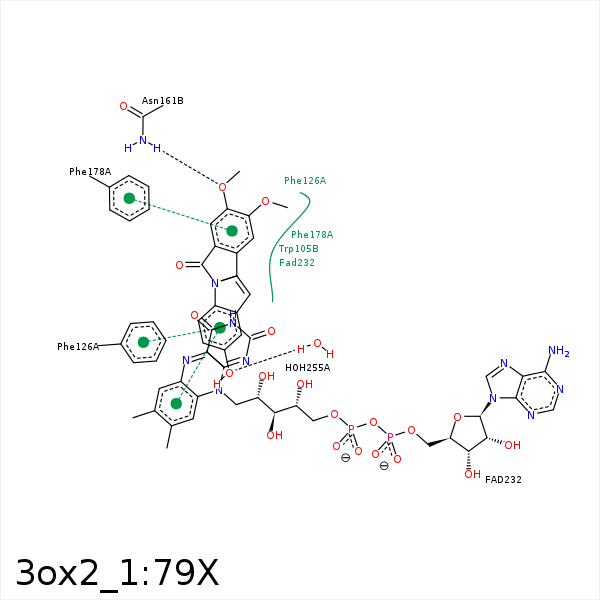


Represent the protein/ligand binding mode, centered on the ligand
Dashed lines represents hydrogen bonds and metal interactions
Green residue labels for amino acids with hydrophobic contacts (green lines) to the ligand
| Ligand | Protein | Interaction | |||
|---|---|---|---|---|---|
| Atom | Atom | Residue | Distance (Å) | Angle (°) | Type |
| C21 | CE3 | TRP- 105 | 4.26 | 0 | Hydrophobic |
| C21 | CE1 | PHE- 106 | 3.39 | 0 | Hydrophobic |
| C2 | CZ | PHE- 126 | 3.21 | 0 | Hydrophobic |
| C23 | SD | MET- 154 | 4.09 | 0 | Hydrophobic |
| O4 | ND2 | ASN- 161 | 2.76 | 148.84 | H-Bond (Protein Donor) |
| C21 | CD1 | PHE- 178 | 3.67 | 0 | Hydrophobic |
| C11 | C1' | FAD- 232 | 4.32 | 0 | Hydrophobic |
| C1 | C7M | FAD- 232 | 3.45 | 0 | Hydrophobic |
| C5 | C8M | FAD- 232 | 3.42 | 0 | Hydrophobic |
| O1 | O | HOH- 255 | 2.78 | 152.36 | H-Bond (Protein Donor) |
