sc-PDB
An Annotated Database of Druggable Binding Sites from the Protein DataBank
An Annotated Database of Druggable Binding Sites from the Protein DataBank
1.600 Å
X-ray
2010-03-03
| Name: | Monomeric sarcosine oxidase |
|---|---|
| ID: | MSOX_BACB0 |
| AC: | P40859 |
| Organism: | Bacillus sp. |
| Reign: | Bacteria |
| TaxID: | 69000 |
| EC Number: | 1.5.3.1 |
| Chain Name: | Percentage of Residues within binding site |
|---|---|
| A | 100 % |
| B-Factor: | 15.381 |
|---|---|
| Number of residues: | 74 |
| Including | |
| Standard Amino Acids: | 67 |
| Non Standard Amino Acids: | 1 |
| Water Molecules: | 6 |
| Cofactors: | |
| Metals: | CL |
| Ligandability | Volume (Å3) |
|---|---|
| 0.750 | 469.125 |
| % Hydrophobic | % Polar |
|---|---|
| 47.48 | 52.52 |
| According to VolSite | |
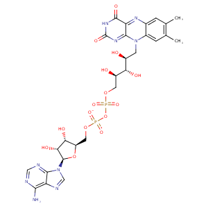
| HET Code: | FAD |
|---|---|
| Formula: | C27H31N9O15P2 |
| Molecular weight: | 783.534 g/mol |
| DrugBank ID: | DB03147 |
| Buried Surface Area: | 79.91 % |
| Polar Surface area: | 381.7 Å2 |
| Number of | |
|---|---|
| H-Bond Acceptors: | 22 |
| H-Bond Donors: | 7 |
| Rings: | 6 |
| Aromatic rings: | 3 |
| Anionic atoms: | 2 |
| Cationic atoms: | 0 |
| Rule of Five Violation: | 3 |
| Rotatable Bonds: | 13 |
| X | Y | Z |
|---|---|---|
| 6.07489 | 37.7521 | 35.4291 |


Represent the protein/ligand binding mode, centered on the ligand
Dashed lines represents hydrogen bonds and metal interactions
Green residue labels for amino acids with hydrophobic contacts (green lines) to the ligand
| Ligand | Protein | Interaction | |||
|---|---|---|---|---|---|
| Atom | Atom | Residue | Distance (Å) | Angle (°) | Type |
| O2A | N | SER- 13 | 2.97 | 173.18 | H-Bond (Protein Donor) |
| O2A | OG | SER- 13 | 2.83 | 165.7 | H-Bond (Protein Donor) |
| O4' | OG | SER- 13 | 2.85 | 159.28 | H-Bond (Ligand Donor) |
| C4' | CB | SER- 13 | 4.12 | 0 | Hydrophobic |
| O2P | N | MET- 14 | 2.99 | 171.98 | H-Bond (Protein Donor) |
| C5' | CE | MET- 14 | 4.27 | 0 | Hydrophobic |
| O3B | OD1 | ASP- 33 | 2.69 | 175.14 | H-Bond (Ligand Donor) |
| O3B | OD2 | ASP- 33 | 3.21 | 120.43 | H-Bond (Ligand Donor) |
| O2B | OD2 | ASP- 33 | 2.81 | 160.31 | H-Bond (Ligand Donor) |
| N3A | N | ALA- 34 | 3.15 | 136.25 | H-Bond (Protein Donor) |
| O3B | NE2 | HIS- 39 | 2.88 | 159.79 | H-Bond (Protein Donor) |
| O2B | NE2 | HIS- 39 | 3.19 | 120.36 | H-Bond (Protein Donor) |
| O1A | N | SER- 43 | 2.85 | 148.45 | H-Bond (Protein Donor) |
| O2' | O | SER- 43 | 2.71 | 137.31 | H-Bond (Ligand Donor) |
| C5' | CB | SER- 43 | 3.99 | 0 | Hydrophobic |
| O2A | ND1 | HIS- 44 | 2.83 | 172.68 | H-Bond (Protein Donor) |
| C6 | CD | ARG- 49 | 4.33 | 0 | Hydrophobic |
| C9A | CD | ARG- 49 | 3.79 | 0 | Hydrophobic |
| O2' | NE | ARG- 49 | 2.93 | 168.94 | H-Bond (Protein Donor) |
| DuAr | CZ | ARG- 49 | 3.74 | 170.09 | Pi/Cation |
| N3 | O | ILE- 50 | 2.98 | 153.75 | H-Bond (Ligand Donor) |
| O4 | N | ILE- 50 | 2.82 | 158.29 | H-Bond (Protein Donor) |
| N6A | O | VAL- 173 | 3.05 | 163.42 | H-Bond (Ligand Donor) |
| N1A | N | VAL- 173 | 2.9 | 169.78 | H-Bond (Protein Donor) |
| C2B | CZ3 | TRP- 204 | 4.23 | 0 | Hydrophobic |
| C7M | CB | GLN- 223 | 4.3 | 0 | Hydrophobic |
| C8M | CB | GLN- 223 | 4.29 | 0 | Hydrophobic |
| C7M | CG1 | VAL- 225 | 3.72 | 0 | Hydrophobic |
| C7M | CZ | TYR- 254 | 4.49 | 0 | Hydrophobic |
| C7M | CB | CYS- 315 | 3.56 | 0 | Hydrophobic |
| C9 | SG | CYS- 315 | 3.89 | 0 | Hydrophobic |
| C8 | SG | CYS- 315 | 3.63 | 0 | Hydrophobic |
| C7M | CE1 | TYR- 317 | 4.44 | 0 | Hydrophobic |
| C8M | CD1 | TYR- 317 | 3.54 | 0 | Hydrophobic |
| C5' | CB | PHE- 342 | 4.35 | 0 | Hydrophobic |
| O3' | N | GLY- 346 | 3.23 | 138.4 | H-Bond (Protein Donor) |
| N1 | N | PHE- 347 | 3.21 | 155.75 | H-Bond (Protein Donor) |
| C2' | CB | PHE- 347 | 3.69 | 0 | Hydrophobic |
| O2 | N | LYS- 348 | 2.86 | 175.43 | H-Bond (Protein Donor) |
| O2 | NZ | LYS- 348 | 2.76 | 142.37 | H-Bond (Protein Donor) |
| O2P | O | HOH- 416 | 2.58 | 161.5 | H-Bond (Protein Donor) |
| O1P | O | HOH- 421 | 2.78 | 140.23 | H-Bond (Protein Donor) |
| O1A | O | HOH- 767 | 2.73 | 179.98 | H-Bond (Protein Donor) |
