sc-PDB
An Annotated Database of Druggable Binding Sites from the Protein DataBank
An Annotated Database of Druggable Binding Sites from the Protein DataBank
2.100 Å
X-ray
2010-01-11
| Name: | Phosphopantetheine adenylyltransferase |
|---|---|
| ID: | COAD_MYCTU |
| AC: | P9WPA5 |
| Organism: | Mycobacterium tuberculosis |
| Reign: | Bacteria |
| TaxID: | 83332 |
| EC Number: | / |
| Chain Name: | Percentage of Residues within binding site |
|---|---|
| A | 100 % |
| B-Factor: | 29.200 |
|---|---|
| Number of residues: | 30 |
| Including | |
| Standard Amino Acids: | 29 |
| Non Standard Amino Acids: | 0 |
| Water Molecules: | 1 |
| Cofactors: | |
| Metals: | |
| Ligandability | Volume (Å3) |
|---|---|
| 0.055 | 411.750 |
| % Hydrophobic | % Polar |
|---|---|
| 33.61 | 66.39 |
| According to VolSite | |
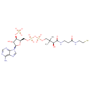
| HET Code: | COA |
|---|---|
| Formula: | C21H32N7O16P3S |
| Molecular weight: | 763.502 g/mol |
| DrugBank ID: | DB01992 |
| Buried Surface Area: | 26.94 % |
| Polar Surface area: | 426.11 Å2 |
| Number of | |
|---|---|
| H-Bond Acceptors: | 21 |
| H-Bond Donors: | 6 |
| Rings: | 3 |
| Aromatic rings: | 2 |
| Anionic atoms: | 4 |
| Cationic atoms: | 0 |
| Rule of Five Violation: | 3 |
| Rotatable Bonds: | 18 |
| X | Y | Z |
|---|---|---|
| 11.4253 | 0.0808095 | 16.8273 |
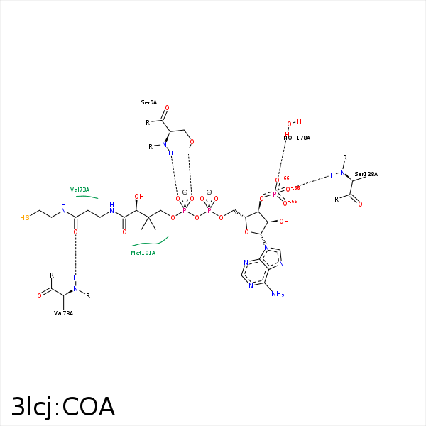


Represent the protein/ligand binding mode, centered on the ligand
Dashed lines represents hydrogen bonds and metal interactions
Green residue labels for amino acids with hydrophobic contacts (green lines) to the ligand
| Ligand | Protein | Interaction | |||
|---|---|---|---|---|---|
| Atom | Atom | Residue | Distance (Å) | Angle (°) | Type |
| O2B | OG | SER- 9 | 3.41 | 124.5 | H-Bond (Ligand Donor) |
| O4A | OG | SER- 9 | 3.3 | 137.28 | H-Bond (Protein Donor) |
| O4A | N | SER- 9 | 2.98 | 152.86 | H-Bond (Protein Donor) |
| O5A | OG | SER- 9 | 2.96 | 155.1 | H-Bond (Protein Donor) |
| C6P | CG2 | VAL- 73 | 4.1 | 0 | Hydrophobic |
| O5P | N | VAL- 73 | 2.99 | 171.02 | H-Bond (Protein Donor) |
| C2P | SD | MET- 101 | 3.87 | 0 | Hydrophobic |
| S1P | CG | MET- 101 | 3.58 | 0 | Hydrophobic |
| CEP | CE | MET- 101 | 3.68 | 0 | Hydrophobic |
| S1P | CG | MET- 104 | 4.24 | 0 | Hydrophobic |
| O8A | N | SER- 128 | 3.29 | 159.34 | H-Bond (Protein Donor) |
| O9A | O | HOH- 178 | 2.55 | 159.06 | H-Bond (Protein Donor) |
