sc-PDB
An Annotated Database of Druggable Binding Sites from the Protein DataBank
An Annotated Database of Druggable Binding Sites from the Protein DataBank
2.050 Å
X-ray
2009-07-23
| Name: | Purine nucleoside phosphorylase |
|---|---|
| ID: | Q9BMI9_SCHMA |
| AC: | Q9BMI9 |
| Organism: | Schistosoma mansoni |
| Reign: | Eukaryota |
| TaxID: | 6183 |
| EC Number: | / |
| Chain Name: | Percentage of Residues within binding site |
|---|---|
| A | 97 % |
| B | 3 % |
| B-Factor: | 29.052 |
|---|---|
| Number of residues: | 35 |
| Including | |
| Standard Amino Acids: | 34 |
| Non Standard Amino Acids: | 0 |
| Water Molecules: | 1 |
| Cofactors: | |
| Metals: | |
| Ligandability | Volume (Å3) |
|---|---|
| 0.811 | 617.625 |
| % Hydrophobic | % Polar |
|---|---|
| 39.89 | 60.11 |
| According to VolSite | |
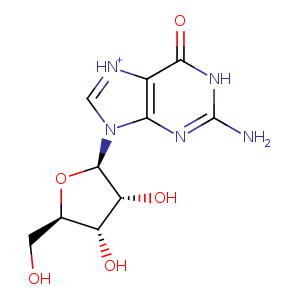
| HET Code: | GMP |
|---|---|
| Formula: | C10H13N5O5 |
| Molecular weight: | 283.241 g/mol |
| DrugBank ID: | DB02857 |
| Buried Surface Area: | 82.16 % |
| Polar Surface area: | 155.22 Å2 |
| Number of | |
|---|---|
| H-Bond Acceptors: | 8 |
| H-Bond Donors: | 5 |
| Rings: | 3 |
| Aromatic rings: | 1 |
| Anionic atoms: | 0 |
| Cationic atoms: | 0 |
| Rule of Five Violation: | 0 |
| Rotatable Bonds: | 2 |
| X | Y | Z |
|---|---|---|
| -18.7356 | 2.08285 | -29.6333 |
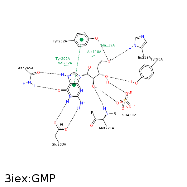


Represent the protein/ligand binding mode, centered on the ligand
Dashed lines represents hydrogen bonds and metal interactions
Green residue labels for amino acids with hydrophobic contacts (green lines) to the ligand
| Ligand | Protein | Interaction | |||
|---|---|---|---|---|---|
| Atom | Atom | Residue | Distance (Å) | Angle (°) | Type |
| C3' | CE2 | TYR- 90 | 4.17 | 0 | Hydrophobic |
| O3' | OH | TYR- 90 | 2.93 | 134.27 | H-Bond (Protein Donor) |
| C1' | CB | ALA- 118 | 3.82 | 0 | Hydrophobic |
| C5' | CD1 | PHE- 161 | 3.57 | 0 | Hydrophobic |
| C4' | CZ | PHE- 161 | 3.81 | 0 | Hydrophobic |
| C3' | CE1 | PHE- 161 | 3.55 | 0 | Hydrophobic |
| C5' | CZ | TYR- 202 | 3.73 | 0 | Hydrophobic |
| N1 | OE2 | GLU- 203 | 3.26 | 124.54 | H-Bond (Ligand Donor) |
| N1 | OE1 | GLU- 203 | 2.7 | 160.29 | H-Bond (Ligand Donor) |
| N2 | OE2 | GLU- 203 | 2.52 | 166.01 | H-Bond (Ligand Donor) |
| C3' | SD | MET- 221 | 3.62 | 0 | Hydrophobic |
| C2' | CG | MET- 221 | 3.67 | 0 | Hydrophobic |
| O2' | N | MET- 221 | 2.89 | 147.7 | H-Bond (Protein Donor) |
| O6 | ND2 | ASN- 245 | 2.9 | 165 | H-Bond (Protein Donor) |
| O5' | ND1 | HIS- 259 | 2.75 | 149.65 | H-Bond (Protein Donor) |
