sc-PDB
An Annotated Database of Druggable Binding Sites from the Protein DataBank
An Annotated Database of Druggable Binding Sites from the Protein DataBank
1.870 Å
X-ray
2009-07-07
| Name: | Putative leucoanthocyanidin reductase 1 |
|---|---|
| ID: | Q4W2K4_VITVI |
| AC: | Q4W2K4 |
| Organism: | Vitis vinifera |
| Reign: | Eukaryota |
| TaxID: | 29760 |
| EC Number: | / |
| Chain Name: | Percentage of Residues within binding site |
|---|---|
| A | 100 % |
| B-Factor: | 18.187 |
|---|---|
| Number of residues: | 41 |
| Including | |
| Standard Amino Acids: | 39 |
| Non Standard Amino Acids: | 0 |
| Water Molecules: | 2 |
| Cofactors: | |
| Metals: | |
| Ligandability | Volume (Å3) |
|---|---|
| 1.061 | 722.250 |
| % Hydrophobic | % Polar |
|---|---|
| 43.46 | 56.54 |
| According to VolSite | |
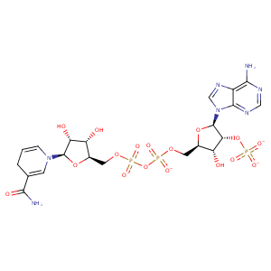
| HET Code: | NDP |
|---|---|
| Formula: | C21H26N7O17P3 |
| Molecular weight: | 741.389 g/mol |
| DrugBank ID: | DB02338 |
| Buried Surface Area: | 51.91 % |
| Polar Surface area: | 404.9 Å2 |
| Number of | |
|---|---|
| H-Bond Acceptors: | 22 |
| H-Bond Donors: | 5 |
| Rings: | 5 |
| Aromatic rings: | 2 |
| Anionic atoms: | 4 |
| Cationic atoms: | 0 |
| Rule of Five Violation: | 2 |
| Rotatable Bonds: | 13 |
| X | Y | Z |
|---|---|---|
| 3.19679 | 8.31746 | 19.053 |
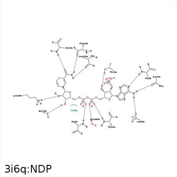


Represent the protein/ligand binding mode, centered on the ligand
Dashed lines represents hydrogen bonds and metal interactions
Green residue labels for amino acids with hydrophobic contacts (green lines) to the ligand
| Ligand | Protein | Interaction | |||
|---|---|---|---|---|---|
| Atom | Atom | Residue | Distance (Å) | Angle (°) | Type |
| O3B | OG1 | THR- 19 | 3.08 | 143.05 | H-Bond (Protein Donor) |
| O2A | N | PHE- 21 | 2.95 | 165.38 | H-Bond (Protein Donor) |
| O2N | N | ILE- 22 | 2.86 | 171.25 | H-Bond (Protein Donor) |
| C1D | CG1 | ILE- 22 | 4.49 | 0 | Hydrophobic |
| C5D | CG1 | ILE- 22 | 4.08 | 0 | Hydrophobic |
| N1A | N | ILE- 69 | 3.01 | 166.47 | H-Bond (Protein Donor) |
| N6A | OD1 | ASN- 70 | 2.99 | 148.48 | H-Bond (Ligand Donor) |
| C1B | CG1 | VAL- 91 | 4.42 | 0 | Hydrophobic |
| N6A | OD2 | ASP- 98 | 2.98 | 149.09 | H-Bond (Ligand Donor) |
| O3D | OG | SER- 118 | 2.87 | 163.02 | H-Bond (Ligand Donor) |
| C4D | CB | PHE- 120 | 4.48 | 0 | Hydrophobic |
| C1D | CB | PHE- 120 | 4.23 | 0 | Hydrophobic |
| N7N | O | PHE- 120 | 2.84 | 161.8 | H-Bond (Ligand Donor) |
| O2D | NZ | LYS- 140 | 2.86 | 157.58 | H-Bond (Protein Donor) |
| N7N | O | ASN- 160 | 2.63 | 147.9 | H-Bond (Ligand Donor) |
| O7N | N | ILE- 162 | 2.99 | 174.42 | H-Bond (Protein Donor) |
| O1A | O | HOH- 459 | 2.7 | 179.99 | H-Bond (Protein Donor) |
