sc-PDB
An Annotated Database of Druggable Binding Sites from the Protein DataBank
An Annotated Database of Druggable Binding Sites from the Protein DataBank
2.200 Å
X-ray
2008-05-07
| Name: | Methionine aminopeptidase |
|---|---|
| ID: | MAP1_ECOLI |
| AC: | P0AE18 |
| Organism: | Escherichia coli |
| Reign: | Bacteria |
| TaxID: | 83333 |
| EC Number: | / |
| Chain Name: | Percentage of Residues within binding site |
|---|---|
| A | 100 % |
| B-Factor: | 18.055 |
|---|---|
| Number of residues: | 30 |
| Including | |
| Standard Amino Acids: | 28 |
| Non Standard Amino Acids: | 2 |
| Water Molecules: | 0 |
| Cofactors: | |
| Metals: | MN MN |
| Ligandability | Volume (Å3) |
|---|---|
| 0.687 | 330.750 |
| % Hydrophobic | % Polar |
|---|---|
| 52.04 | 47.96 |
| According to VolSite | |
| HET Code: | W29 |
|---|---|
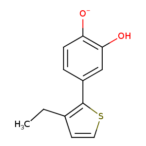
| Formula: | C12H12O2S |
| Molecular weight: | 220.287 g/mol |
| DrugBank ID: | DB08718 |
| Buried Surface Area: | 69.04 % |
| Polar Surface area: | 68.7 Å2 |
| Number of | |
|---|---|
| H-Bond Acceptors: | 2 |
| H-Bond Donors: | 2 |
| Rings: | 2 |
| Aromatic rings: | 2 |
| Anionic atoms: | 0 |
| Cationic atoms: | 0 |
| Rule of Five Violation: | 0 |
| Rotatable Bonds: | 2 |
| X | Y | Z |
|---|---|---|
| -2.94347 | 13.7465 | 8.91013 |
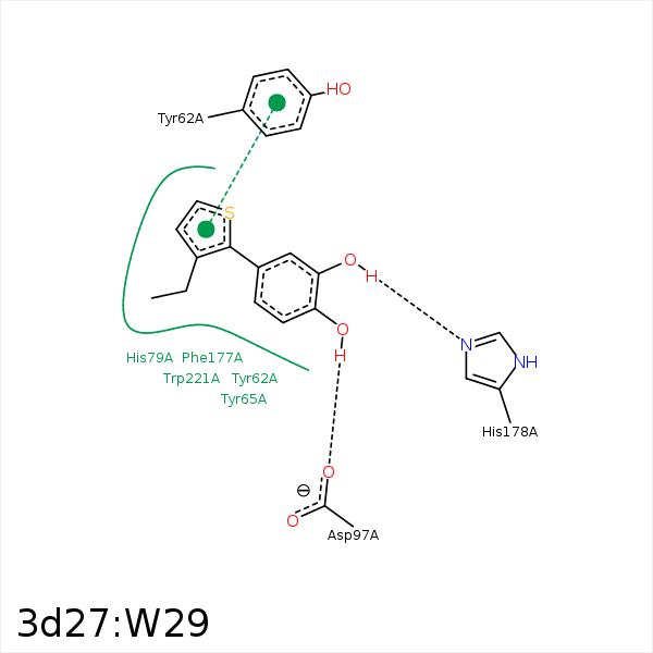


Represent the protein/ligand binding mode, centered on the ligand
Dashed lines represents hydrogen bonds and metal interactions
Green residue labels for amino acids with hydrophobic contacts (green lines) to the ligand
| Ligand | Protein | Interaction | |||
|---|---|---|---|---|---|
| Atom | Atom | Residue | Distance (Å) | Angle (°) | Type |
| O2 | MN | MN- 1 | 2.36 | 0 | Metal Acceptor |
| O1 | MN | MN- 1 | 2.42 | 0 | Metal Acceptor |
| O2 | MN | MN- 2 | 2.01 | 0 | Metal Acceptor |
| C11 | CB | CYS- 59 | 4.47 | 0 | Hydrophobic |
| C11 | CD2 | TYR- 62 | 3.68 | 0 | Hydrophobic |
| C11 | CB | TYR- 65 | 4.23 | 0 | Hydrophobic |
| C12 | CG | TYR- 65 | 3.55 | 0 | Hydrophobic |
| C04 | SG | CYS- 70 | 4.32 | 0 | Hydrophobic |
| C12 | SG | CYS- 70 | 3.41 | 0 | Hydrophobic |
| O1 | NE2 | HIS- 178 | 2.52 | 135.44 | H-Bond (Ligand Donor) |
