sc-PDB
An Annotated Database of Druggable Binding Sites from the Protein DataBank
An Annotated Database of Druggable Binding Sites from the Protein DataBank
2.600 Å
X-ray
2009-02-09
| Name: | Thioredoxin reductase 1, cytoplasmic |
|---|---|
| ID: | TRXR1_HUMAN |
| AC: | Q16881 |
| Organism: | Homo sapiens |
| Reign: | Eukaryota |
| TaxID: | 9606 |
| EC Number: | 1.8.1.9 |
| Chain Name: | Percentage of Residues within binding site |
|---|---|
| C | 94 % |
| D | 6 % |
| B-Factor: | 51.266 |
|---|---|
| Number of residues: | 68 |
| Including | |
| Standard Amino Acids: | 66 |
| Non Standard Amino Acids: | 1 |
| Water Molecules: | 1 |
| Cofactors: | NAP |
| Metals: | |
| Ligandability | Volume (Å3) |
|---|---|
| 1.052 | 938.250 |
| % Hydrophobic | % Polar |
|---|---|
| 44.24 | 55.76 |
| According to VolSite | |
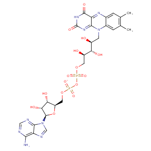
| HET Code: | FAD |
|---|---|
| Formula: | C27H31N9O15P2 |
| Molecular weight: | 783.534 g/mol |
| DrugBank ID: | DB03147 |
| Buried Surface Area: | 77.29 % |
| Polar Surface area: | 381.7 Å2 |
| Number of | |
|---|---|
| H-Bond Acceptors: | 22 |
| H-Bond Donors: | 7 |
| Rings: | 6 |
| Aromatic rings: | 3 |
| Anionic atoms: | 2 |
| Cationic atoms: | 0 |
| Rule of Five Violation: | 3 |
| Rotatable Bonds: | 13 |
| X | Y | Z |
|---|---|---|
| -19.7415 | 50.2544 | -45.9888 |
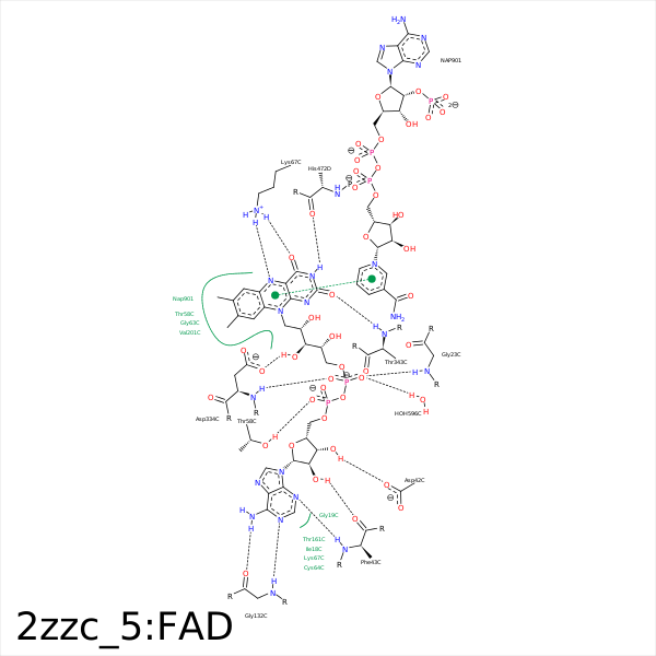


Represent the protein/ligand binding mode, centered on the ligand
Dashed lines represents hydrogen bonds and metal interactions
Green residue labels for amino acids with hydrophobic contacts (green lines) to the ligand
| Ligand | Protein | Interaction | |||
|---|---|---|---|---|---|
| Atom | Atom | Residue | Distance (Å) | Angle (°) | Type |
| O1A | N | SER- 22 | 3.49 | 154.54 | H-Bond (Protein Donor) |
| O1P | N | GLY- 23 | 2.99 | 139.77 | H-Bond (Protein Donor) |
| O3B | OD1 | ASP- 42 | 3.06 | 163.26 | H-Bond (Ligand Donor) |
| O2B | O | PHE- 43 | 2.86 | 154.04 | H-Bond (Ligand Donor) |
| N3A | N | PHE- 43 | 3.06 | 147.13 | H-Bond (Protein Donor) |
| O1A | N | THR- 58 | 3.39 | 150.91 | H-Bond (Protein Donor) |
| O2A | N | THR- 58 | 3.44 | 146.87 | H-Bond (Protein Donor) |
| O2A | OG1 | THR- 58 | 2.59 | 170.39 | H-Bond (Protein Donor) |
| C8M | CG2 | THR- 58 | 3.83 | 0 | Hydrophobic |
| C2' | SG | CYS- 64 | 4.24 | 0 | Hydrophobic |
| O4 | NZ | LYS- 67 | 2.81 | 144.97 | H-Bond (Protein Donor) |
| N5 | NZ | LYS- 67 | 3.12 | 134.98 | H-Bond (Protein Donor) |
| C6 | CB | LYS- 67 | 4.45 | 0 | Hydrophobic |
| N6A | O | GLY- 132 | 2.94 | 162.57 | H-Bond (Ligand Donor) |
| N1A | N | GLY- 132 | 2.89 | 165.45 | H-Bond (Protein Donor) |
| C7M | CB | SER- 180 | 3.85 | 0 | Hydrophobic |
| C7M | CE2 | PHE- 184 | 3.8 | 0 | Hydrophobic |
| C7M | CG2 | VAL- 201 | 3.99 | 0 | Hydrophobic |
| C8M | CD | ARG- 293 | 4.31 | 0 | Hydrophobic |
| O3' | OD2 | ASP- 334 | 3.35 | 128.04 | H-Bond (Ligand Donor) |
| O3' | OD1 | ASP- 334 | 2.69 | 173.08 | H-Bond (Ligand Donor) |
| O2P | N | ASP- 334 | 3.01 | 156.09 | H-Bond (Protein Donor) |
| N1 | N | THR- 343 | 3.5 | 178.43 | H-Bond (Protein Donor) |
| O2 | N | THR- 343 | 3 | 127.35 | H-Bond (Protein Donor) |
| C2' | CB | THR- 343 | 4.42 | 0 | Hydrophobic |
| C4' | CB | THR- 343 | 4.39 | 0 | Hydrophobic |
| C5' | CB | ALA- 346 | 4.31 | 0 | Hydrophobic |
| N3 | O | HIS- 472 | 2.89 | 151.75 | H-Bond (Ligand Donor) |
