sc-PDB
An Annotated Database of Druggable Binding Sites from the Protein DataBank
An Annotated Database of Druggable Binding Sites from the Protein DataBank
2.300 Å
X-ray
2011-04-11
| Name: | Putrescine oxidase |
|---|---|
| ID: | B0F9F6_RHOER |
| AC: | B0F9F6 |
| Organism: | Rhodococcus erythropolis |
| Reign: | Bacteria |
| TaxID: | 1833 |
| EC Number: | / |
| Chain Name: | Percentage of Residues within binding site |
|---|---|
| B | 100 % |
| B-Factor: | 11.173 |
|---|---|
| Number of residues: | 71 |
| Including | |
| Standard Amino Acids: | 64 |
| Non Standard Amino Acids: | 0 |
| Water Molecules: | 7 |
| Cofactors: | |
| Metals: | |
| Ligandability | Volume (Å3) |
|---|---|
| 1.141 | 560.250 |
| % Hydrophobic | % Polar |
|---|---|
| 57.83 | 42.17 |
| According to VolSite | |
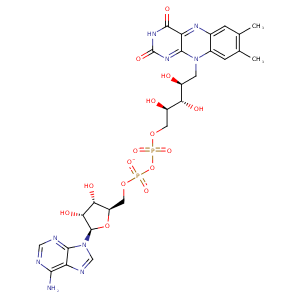
| HET Code: | FAD |
|---|---|
| Formula: | C27H31N9O15P2 |
| Molecular weight: | 783.534 g/mol |
| DrugBank ID: | DB03147 |
| Buried Surface Area: | 79.58 % |
| Polar Surface area: | 381.7 Å2 |
| Number of | |
|---|---|
| H-Bond Acceptors: | 22 |
| H-Bond Donors: | 7 |
| Rings: | 6 |
| Aromatic rings: | 3 |
| Anionic atoms: | 2 |
| Cationic atoms: | 0 |
| Rule of Five Violation: | 3 |
| Rotatable Bonds: | 13 |
| X | Y | Z |
|---|---|---|
| -22.1065 | 32.2646 | -11.2593 |


Represent the protein/ligand binding mode, centered on the ligand
Dashed lines represents hydrogen bonds and metal interactions
Green residue labels for amino acids with hydrophobic contacts (green lines) to the ligand
| Ligand | Protein | Interaction | |||
|---|---|---|---|---|---|
| Atom | Atom | Residue | Distance (Å) | Angle (°) | Type |
| C4' | CG | PRO- 15 | 4.07 | 0 | Hydrophobic |
| O1P | N | SER- 16 | 2.71 | 153.01 | H-Bond (Protein Donor) |
| O3B | OE1 | GLU- 35 | 2.56 | 155.49 | H-Bond (Ligand Donor) |
| O3B | OE2 | GLU- 35 | 3.29 | 122.17 | H-Bond (Ligand Donor) |
| O2B | OE2 | GLU- 35 | 2.63 | 152.51 | H-Bond (Ligand Donor) |
| N3A | N | ALA- 36 | 3.1 | 142.6 | H-Bond (Protein Donor) |
| O2B | NH1 | ARG- 37 | 2.89 | 126.39 | H-Bond (Protein Donor) |
| O1A | NH2 | ARG- 43 | 3.28 | 130.13 | H-Bond (Protein Donor) |
| O1A | NE | ARG- 43 | 2.68 | 168.36 | H-Bond (Protein Donor) |
| O2A | N | ARG- 43 | 2.89 | 165.28 | H-Bond (Protein Donor) |
| O3P | NH2 | ARG- 43 | 3.1 | 140.86 | H-Bond (Protein Donor) |
| O1A | CZ | ARG- 43 | 3.4 | 0 | Ionic (Protein Cationic) |
| C8M | CG | ARG- 43 | 3.98 | 0 | Hydrophobic |
| C9 | CB | ARG- 43 | 4.25 | 0 | Hydrophobic |
| C3' | CB | ARG- 43 | 4.15 | 0 | Hydrophobic |
| O4 | N | GLN- 59 | 3.11 | 163 | H-Bond (Protein Donor) |
| N3 | O | TRP- 60 | 3.12 | 168.14 | H-Bond (Ligand Donor) |
| O4 | N | TRP- 60 | 2.97 | 170.39 | H-Bond (Protein Donor) |
| N6A | O | VAL- 235 | 3.24 | 163.46 | H-Bond (Ligand Donor) |
| N1A | N | VAL- 235 | 2.98 | 174.71 | H-Bond (Protein Donor) |
| C1B | CG1 | VAL- 264 | 4.41 | 0 | Hydrophobic |
| C7M | CD | LYS- 296 | 4.08 | 0 | Hydrophobic |
| C7M | CE2 | TRP- 385 | 4.49 | 0 | Hydrophobic |
| C8M | CE2 | TRP- 385 | 3.69 | 0 | Hydrophobic |
| C2B | CB | TRP- 390 | 4 | 0 | Hydrophobic |
| C7M | CB | ALA- 394 | 4.32 | 0 | Hydrophobic |
| C8M | CB | ALA- 394 | 3.61 | 0 | Hydrophobic |
| C7M | CD1 | TYR- 395 | 4.12 | 0 | Hydrophobic |
| C1' | CB | TYR- 395 | 4.39 | 0 | Hydrophobic |
| C6 | CD1 | TYR- 395 | 3.17 | 0 | Hydrophobic |
| C9A | CB | TYR- 395 | 4.04 | 0 | Hydrophobic |
| O3' | OG | SER- 423 | 2.89 | 155.38 | H-Bond (Protein Donor) |
| O2P | N | SER- 423 | 2.82 | 152.09 | H-Bond (Protein Donor) |
| C5' | CB | SER- 423 | 3.99 | 0 | Hydrophobic |
| N1 | N | VAL- 433 | 3.48 | 149.96 | H-Bond (Protein Donor) |
| O2 | N | VAL- 433 | 2.88 | 148.48 | H-Bond (Protein Donor) |
| C2' | CG2 | VAL- 433 | 3.99 | 0 | Hydrophobic |
| C4' | CG2 | VAL- 433 | 4.28 | 0 | Hydrophobic |
| C5' | CB | ALA- 436 | 4.08 | 0 | Hydrophobic |
| O1P | O | HOH- 2009 | 2.83 | 179.97 | H-Bond (Protein Donor) |
| O3B | O | HOH- 2023 | 2.87 | 179.96 | H-Bond (Protein Donor) |
