sc-PDB
An Annotated Database of Druggable Binding Sites from the Protein DataBank
An Annotated Database of Druggable Binding Sites from the Protein DataBank
1.700 Å
X-ray
2010-06-25
| Name: | Genome polyprotein |
|---|---|
| ID: | POLG_HCVH |
| AC: | P27958 |
| Organism: | Hepatitis C virus genotype 1a |
| Reign: | Viruses |
| TaxID: | 11108 |
| EC Number: | 2.7.7.48 |
| Chain Name: | Percentage of Residues within binding site |
|---|---|
| A | 100 % |
| B-Factor: | 24.034 |
|---|---|
| Number of residues: | 29 |
| Including | |
| Standard Amino Acids: | 26 |
| Non Standard Amino Acids: | 3 |
| Water Molecules: | 0 |
| Cofactors: | GTP |
| Metals: | MG MG |
| Ligandability | Volume (Å3) |
|---|---|
| 0.278 | 2460.375 |
| % Hydrophobic | % Polar |
|---|---|
| 30.59 | 69.41 |
| According to VolSite | |
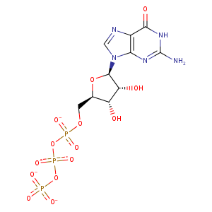
| HET Code: | GTP |
|---|---|
| Formula: | C10H12N5O14P3 |
| Molecular weight: | 519.149 g/mol |
| DrugBank ID: | DB04137 |
| Buried Surface Area: | 46.9 % |
| Polar Surface area: | 335.56 Å2 |
| Number of | |
|---|---|
| H-Bond Acceptors: | 17 |
| H-Bond Donors: | 4 |
| Rings: | 3 |
| Aromatic rings: | 1 |
| Anionic atoms: | 4 |
| Cationic atoms: | 0 |
| Rule of Five Violation: | 2 |
| Rotatable Bonds: | 8 |
| X | Y | Z |
|---|---|---|
| 7.43697 | 0.388312 | 15.5151 |
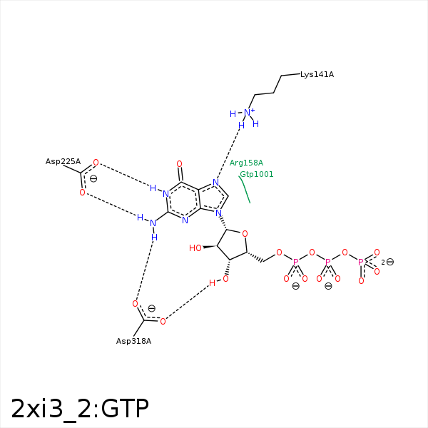


Represent the protein/ligand binding mode, centered on the ligand
Dashed lines represents hydrogen bonds and metal interactions
Green residue labels for amino acids with hydrophobic contacts (green lines) to the ligand
| Ligand | Protein | Interaction | |||
|---|---|---|---|---|---|
| Atom | Atom | Residue | Distance (Å) | Angle (°) | Type |
| O1B | NH1 | ARG- 48 | 3.18 | 133.43 | H-Bond (Protein Donor) |
| O1G | NZ | LYS- 51 | 3.88 | 0 | Ionic (Protein Cationic) |
| N7 | NZ | LYS- 141 | 2.96 | 163.24 | H-Bond (Protein Donor) |
| N1 | OD2 | ASP- 225 | 2.92 | 162.31 | H-Bond (Ligand Donor) |
| N2 | OD1 | ASP- 225 | 2.93 | 174.44 | H-Bond (Ligand Donor) |
| O3' | OD1 | ASP- 318 | 2.72 | 164.42 | H-Bond (Ligand Donor) |
| N2 | OD2 | ASP- 318 | 2.87 | 171.56 | H-Bond (Ligand Donor) |
| O2A | MG | MG- 1005 | 2.5 | 0 | Metal Acceptor |
