sc-PDB
An Annotated Database of Druggable Binding Sites from the Protein DataBank
An Annotated Database of Druggable Binding Sites from the Protein DataBank
2.200 Å
X-ray
2008-08-08
| Name: | AcsD |
|---|---|
| ID: | Q93AT8_DICCH |
| AC: | Q93AT8 |
| Organism: | Dickeya chrysanthemi |
| Reign: | Bacteria |
| TaxID: | 556 |
| EC Number: | / |
| Chain Name: | Percentage of Residues within binding site |
|---|---|
| B | 100 % |
| B-Factor: | 11.601 |
|---|---|
| Number of residues: | 42 |
| Including | |
| Standard Amino Acids: | 36 |
| Non Standard Amino Acids: | 1 |
| Water Molecules: | 5 |
| Cofactors: | |
| Metals: | MG |
| Ligandability | Volume (Å3) |
|---|---|
| 0.781 | 1221.750 |
| % Hydrophobic | % Polar |
|---|---|
| 33.70 | 66.30 |
| According to VolSite | |
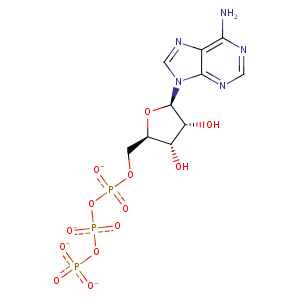
| HET Code: | ATP |
|---|---|
| Formula: | C10H12N5O13P3 |
| Molecular weight: | 503.149 g/mol |
| DrugBank ID: | DB00171 |
| Buried Surface Area: | 69.02 % |
| Polar Surface area: | 319.88 Å2 |
| Number of | |
|---|---|
| H-Bond Acceptors: | 17 |
| H-Bond Donors: | 3 |
| Rings: | 3 |
| Aromatic rings: | 2 |
| Anionic atoms: | 4 |
| Cationic atoms: | 0 |
| Rule of Five Violation: | 2 |
| Rotatable Bonds: | 8 |
| X | Y | Z |
|---|---|---|
| -20.1331 | -11.3503 | -2.22526 |
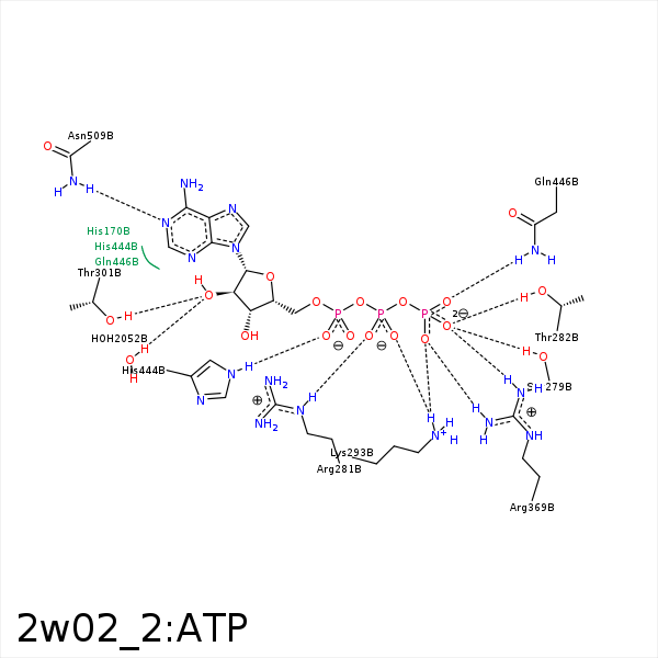


Represent the protein/ligand binding mode, centered on the ligand
Dashed lines represents hydrogen bonds and metal interactions
Green residue labels for amino acids with hydrophobic contacts (green lines) to the ligand
| Ligand | Protein | Interaction | |||
|---|---|---|---|---|---|
| Atom | Atom | Residue | Distance (Å) | Angle (°) | Type |
| O1G | OG | SER- 279 | 2.9 | 162.77 | H-Bond (Protein Donor) |
| O3B | OG | SER- 279 | 3.26 | 123.36 | H-Bond (Protein Donor) |
| O2B | NE | ARG- 281 | 2.76 | 166.14 | H-Bond (Protein Donor) |
| O2B | NH2 | ARG- 281 | 3.24 | 133.93 | H-Bond (Protein Donor) |
| O2B | CZ | ARG- 281 | 3.43 | 0 | Ionic (Protein Cationic) |
| O1G | OG1 | THR- 282 | 2.74 | 161.1 | H-Bond (Protein Donor) |
| O2G | NZ | LYS- 293 | 2.84 | 142.09 | H-Bond (Protein Donor) |
| O1B | NZ | LYS- 293 | 2.9 | 126.24 | H-Bond (Protein Donor) |
| O2G | NZ | LYS- 293 | 2.84 | 0 | Ionic (Protein Cationic) |
| O1B | NZ | LYS- 293 | 2.9 | 0 | Ionic (Protein Cationic) |
| C2' | CG2 | THR- 301 | 4.32 | 0 | Hydrophobic |
| O2' | OG1 | THR- 301 | 2.85 | 160.49 | H-Bond (Protein Donor) |
| O1G | NH1 | ARG- 369 | 3.03 | 174.96 | H-Bond (Protein Donor) |
| O2G | NH2 | ARG- 369 | 2.65 | 174.5 | H-Bond (Protein Donor) |
| O1G | CZ | ARG- 369 | 3.91 | 0 | Ionic (Protein Cationic) |
| O2G | CZ | ARG- 369 | 3.51 | 0 | Ionic (Protein Cationic) |
| O1A | NE2 | HIS- 444 | 2.92 | 131.14 | H-Bond (Protein Donor) |
| O3G | NE2 | GLN- 446 | 3.17 | 147.22 | H-Bond (Protein Donor) |
| C1' | CG | GLN- 446 | 4.26 | 0 | Hydrophobic |
| C4' | CG | GLN- 446 | 4.11 | 0 | Hydrophobic |
| N6 | OD1 | ASN- 509 | 3.45 | 144.21 | H-Bond (Ligand Donor) |
| N1 | ND2 | ASN- 509 | 3.11 | 159.57 | H-Bond (Protein Donor) |
| O3G | MG | MG- 1589 | 2.04 | 0 | Metal Acceptor |
| O1A | MG | MG- 1589 | 2.45 | 0 | Metal Acceptor |
| O3A | MG | MG- 1589 | 2.51 | 0 | Metal Acceptor |
| O2' | O | HOH- 2052 | 3.08 | 179.97 | H-Bond (Protein Donor) |
