sc-PDB
An Annotated Database of Druggable Binding Sites from the Protein DataBank
An Annotated Database of Druggable Binding Sites from the Protein DataBank
2.250 Å
X-ray
2007-10-01
| Name: | Lysine-specific demethylase 4A |
|---|---|
| ID: | KDM4A_HUMAN |
| AC: | O75164 |
| Organism: | Homo sapiens |
| Reign: | Eukaryota |
| TaxID: | 9606 |
| EC Number: | 1.14.11 |
| Chain Name: | Percentage of Residues within binding site |
|---|---|
| B | 100 % |
| B-Factor: | 39.895 |
|---|---|
| Number of residues: | 22 |
| Including | |
| Standard Amino Acids: | 22 |
| Non Standard Amino Acids: | 0 |
| Water Molecules: | 0 |
| Cofactors: | |
| Metals: | |
| Ligandability | Volume (Å3) |
|---|---|
| 0.467 | 847.125 |
| % Hydrophobic | % Polar |
|---|---|
| 35.46 | 64.54 |
| According to VolSite | |
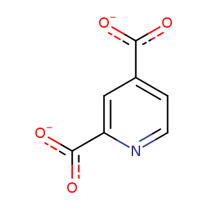
| HET Code: | PD2 |
|---|---|
| Formula: | C7H3NO4 |
| Molecular weight: | 165.103 g/mol |
| DrugBank ID: | - |
| Buried Surface Area: | 70.14 % |
| Polar Surface area: | 93.15 Å2 |
| Number of | |
|---|---|
| H-Bond Acceptors: | 5 |
| H-Bond Donors: | 0 |
| Rings: | 1 |
| Aromatic rings: | 1 |
| Anionic atoms: | 2 |
| Cationic atoms: | 0 |
| Rule of Five Violation: | 0 |
| Rotatable Bonds: | 2 |
| X | Y | Z |
|---|---|---|
| 8.02058 | -55.4143 | 2.50983 |
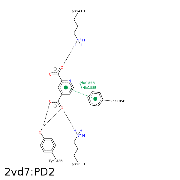


Represent the protein/ligand binding mode, centered on the ligand
Dashed lines represents hydrogen bonds and metal interactions
Green residue labels for amino acids with hydrophobic contacts (green lines) to the ligand
| Ligand | Protein | Interaction | |||
|---|---|---|---|---|---|
| Atom | Atom | Residue | Distance (Å) | Angle (°) | Type |
| O42 | OH | TYR- 132 | 3.04 | 128.14 | H-Bond (Protein Donor) |
| O41 | OH | TYR- 132 | 2.5 | 132.26 | H-Bond (Protein Donor) |
| C4 | CB | PHE- 185 | 3.89 | 0 | Hydrophobic |
| O42 | NZ | LYS- 206 | 2.71 | 170.72 | H-Bond (Protein Donor) |
| O42 | NZ | LYS- 206 | 2.71 | 0 | Ionic (Protein Cationic) |
| O21 | NZ | LYS- 241 | 3.41 | 128.5 | H-Bond (Protein Donor) |
| O22 | NZ | LYS- 241 | 2.86 | 177.23 | H-Bond (Protein Donor) |
| O21 | NZ | LYS- 241 | 3.41 | 0 | Ionic (Protein Cationic) |
| O22 | NZ | LYS- 241 | 2.86 | 0 | Ionic (Protein Cationic) |
