sc-PDB
An Annotated Database of Druggable Binding Sites from the Protein DataBank
An Annotated Database of Druggable Binding Sites from the Protein DataBank
2.510 Å
X-ray
2007-09-06
| Min | Mean | Median | Standard Deviation | Max | Count | |
|---|---|---|---|---|---|---|
| pChEMBL: | 6.080 | 6.080 | 6.080 | 0.000 | 6.080 | 1 |
| Name: | Glutathione S-transferase A1 |
|---|---|
| ID: | GSTA1_HUMAN |
| AC: | P08263 |
| Organism: | Homo sapiens |
| Reign: | Eukaryota |
| TaxID: | 9606 |
| EC Number: | 2.5.1.18 |
| Chain Name: | Percentage of Residues within binding site |
|---|---|
| A | 84 % |
| B | 16 % |
| B-Factor: | 18.405 |
|---|---|
| Number of residues: | 34 |
| Including | |
| Standard Amino Acids: | 32 |
| Non Standard Amino Acids: | 0 |
| Water Molecules: | 2 |
| Cofactors: | |
| Metals: | |
| Ligandability | Volume (Å3) |
|---|---|
| 0.155 | 415.125 |
| % Hydrophobic | % Polar |
|---|---|
| 49.59 | 50.41 |
| According to VolSite | |
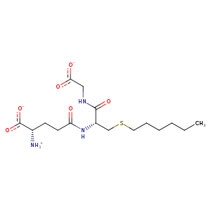
| HET Code: | GTX |
|---|---|
| Formula: | C16H28N3O6S |
| Molecular weight: | 390.475 g/mol |
| DrugBank ID: | - |
| Buried Surface Area: | 58.41 % |
| Polar Surface area: | 191.4 Å2 |
| Number of | |
|---|---|
| H-Bond Acceptors: | 7 |
| H-Bond Donors: | 3 |
| Rings: | 0 |
| Aromatic rings: | 0 |
| Anionic atoms: | 2 |
| Cationic atoms: | 1 |
| Rule of Five Violation: | 0 |
| Rotatable Bonds: | 15 |
| X | Y | Z |
|---|---|---|
| 30.8648 | 9.85573 | 14.5118 |
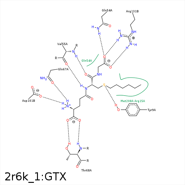


Represent the protein/ligand binding mode, centered on the ligand
Dashed lines represents hydrogen bonds and metal interactions
Green residue labels for amino acids with hydrophobic contacts (green lines) to the ligand
| Ligand | Protein | Interaction | |||
|---|---|---|---|---|---|
| Atom | Atom | Residue | Distance (Å) | Angle (°) | Type |
| SG2 | CE1 | TYR- 9 | 3.68 | 0 | Hydrophobic |
| CB1 | CD | ARG- 15 | 4.06 | 0 | Hydrophobic |
| C2S | CG | ARG- 15 | 3.83 | 0 | Hydrophobic |
| O31 | CZ | ARG- 45 | 3.88 | 0 | Ionic (Protein Cationic) |
| CG1 | CB | GLN- 54 | 4.03 | 0 | Hydrophobic |
| O32 | NE2 | GLN- 54 | 3.25 | 169.15 | H-Bond (Protein Donor) |
| N2 | O | VAL- 55 | 2.54 | 167.23 | H-Bond (Ligand Donor) |
| O2 | N | VAL- 55 | 3.16 | 132.79 | H-Bond (Protein Donor) |
| N1 | OE1 | GLN- 67 | 2.58 | 135.4 | H-Bond (Ligand Donor) |
| O11 | N | THR- 68 | 2.98 | 154.75 | H-Bond (Protein Donor) |
| O12 | N | THR- 68 | 3.43 | 149.98 | H-Bond (Protein Donor) |
| O12 | OG1 | THR- 68 | 2.72 | 165.97 | H-Bond (Protein Donor) |
| N1 | OD1 | ASP- 101 | 3.06 | 120.74 | H-Bond (Ligand Donor) |
| N1 | OD2 | ASP- 101 | 2.55 | 152.8 | H-Bond (Ligand Donor) |
| N1 | OD1 | ASP- 101 | 3.06 | 0 | Ionic (Ligand Cationic) |
| N1 | OD2 | ASP- 101 | 2.55 | 0 | Ionic (Ligand Cationic) |
| C5S | CG2 | VAL- 111 | 4.07 | 0 | Hydrophobic |
| C6S | CB | VAL- 111 | 4.49 | 0 | Hydrophobic |
| O31 | NH1 | ARG- 131 | 3.22 | 136.55 | H-Bond (Protein Donor) |
| O31 | NH2 | ARG- 131 | 2.95 | 149.83 | H-Bond (Protein Donor) |
| O32 | NH1 | ARG- 131 | 2.79 | 148.86 | H-Bond (Protein Donor) |
| O31 | CZ | ARG- 131 | 3.52 | 0 | Ionic (Protein Cationic) |
| O32 | CZ | ARG- 131 | 3.81 | 0 | Ionic (Protein Cationic) |
| C6S | CE | MET- 208 | 4.08 | 0 | Hydrophobic |
| C6S | CD1 | LEU- 213 | 4.42 | 0 | Hydrophobic |
| CB2 | CE1 | PHE- 220 | 3.46 | 0 | Hydrophobic |
| C5S | CZ | PHE- 222 | 3.83 | 0 | Hydrophobic |
