sc-PDB
An Annotated Database of Druggable Binding Sites from the Protein DataBank
An Annotated Database of Druggable Binding Sites from the Protein DataBank
2.450 Å
X-ray
2006-10-26
| Name: | (3,5-dihydroxyphenyl)acetyl-CoA 1,2-dioxygenase |
|---|---|
| ID: | DPGC_STRTO |
| AC: | Q8KLK7 |
| Organism: | Streptomyces toyocaensis |
| Reign: | Bacteria |
| TaxID: | 55952 |
| EC Number: | / |
| Chain Name: | Percentage of Residues within binding site |
|---|---|
| B | 100 % |
| B-Factor: | 32.721 |
|---|---|
| Number of residues: | 40 |
| Including | |
| Standard Amino Acids: | 38 |
| Non Standard Amino Acids: | 0 |
| Water Molecules: | 2 |
| Cofactors: | |
| Metals: | |
| Ligandability | Volume (Å3) |
|---|---|
| 1.532 | 486.000 |
| % Hydrophobic | % Polar |
|---|---|
| 68.75 | 31.25 |
| According to VolSite | |
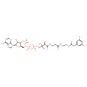
| HET Code: | YE1 |
|---|---|
| Formula: | C29H39N8O19P3 |
| Molecular weight: | 896.583 g/mol |
| DrugBank ID: | - |
| Buried Surface Area: | 57.61 % |
| Polar Surface area: | 456.87 Å2 |
| Number of | |
|---|---|
| H-Bond Acceptors: | 23 |
| H-Bond Donors: | 8 |
| Rings: | 4 |
| Aromatic rings: | 3 |
| Anionic atoms: | 4 |
| Cationic atoms: | 0 |
| Rule of Five Violation: | 3 |
| Rotatable Bonds: | 21 |
| X | Y | Z |
|---|---|---|
| -72.8521 | 31.8405 | -21.5497 |
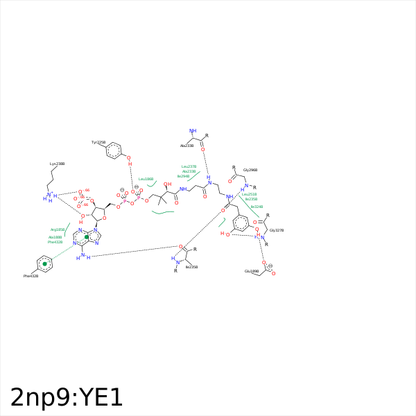


Represent the protein/ligand binding mode, centered on the ligand
Dashed lines represents hydrogen bonds and metal interactions
Green residue labels for amino acids with hydrophobic contacts (green lines) to the ligand
| Ligand | Protein | Interaction | |||
|---|---|---|---|---|---|
| Atom | Atom | Residue | Distance (Å) | Angle (°) | Type |
| C1' | CB | ARG- 185 | 4.37 | 0 | Hydrophobic |
| C12 | CD2 | LEU- 186 | 3.99 | 0 | Hydrophobic |
| C13 | CD2 | LEU- 186 | 4.03 | 0 | Hydrophobic |
| C5' | CD2 | LEU- 186 | 3.75 | 0 | Hydrophobic |
| OAL | OE1 | GLU- 189 | 2.62 | 151.72 | H-Bond (Ligand Donor) |
| O8A | NE2 | HIS- 222 | 3.41 | 162.15 | H-Bond (Protein Donor) |
| O2A | CZ | ARG- 224 | 3.48 | 0 | Ionic (Protein Cationic) |
| O5A | OH | TYR- 225 | 2.57 | 146.38 | H-Bond (Protein Donor) |
| C13 | CB | ALA- 233 | 4.43 | 0 | Hydrophobic |
| N4P | O | ALA- 233 | 3.05 | 144.88 | H-Bond (Ligand Donor) |
| N6A | O | ILE- 235 | 3.05 | 153.51 | H-Bond (Ligand Donor) |
| OAD | N | ILE- 235 | 2.93 | 173.25 | H-Bond (Protein Donor) |
| CAC | CG2 | ILE- 235 | 3.92 | 0 | Hydrophobic |
| CAE | CB | ILE- 235 | 3.54 | 0 | Hydrophobic |
| N1A | N | LEU- 237 | 3.37 | 146.07 | H-Bond (Protein Donor) |
| C6P | CD1 | LEU- 237 | 4.15 | 0 | Hydrophobic |
| O2' | NZ | LYS- 238 | 3.17 | 138.77 | H-Bond (Protein Donor) |
| O9A | NZ | LYS- 238 | 2.87 | 163.2 | H-Bond (Protein Donor) |
| O7A | NZ | LYS- 238 | 3.88 | 0 | Ionic (Protein Cationic) |
| O9A | NZ | LYS- 238 | 2.87 | 0 | Ionic (Protein Cationic) |
| CAH | CD2 | LEU- 251 | 3.83 | 0 | Hydrophobic |
| CAI | CD | ARG- 254 | 3.91 | 0 | Hydrophobic |
| C13 | CZ | PHE- 292 | 3.61 | 0 | Hydrophobic |
| C13 | CD1 | ILE- 294 | 3.95 | 0 | Hydrophobic |
| C6P | CG2 | ILE- 294 | 4.24 | 0 | Hydrophobic |
| OAD | N | GLY- 296 | 2.95 | 168.03 | H-Bond (Protein Donor) |
| CAC | CG1 | ILE- 324 | 3.67 | 0 | Hydrophobic |
| CAG | CD1 | ILE- 324 | 3.28 | 0 | Hydrophobic |
| OAK | N | GLY- 327 | 2.91 | 121.1 | H-Bond (Protein Donor) |
| OAK | NE2 | GLN- 416 | 3.18 | 123.66 | H-Bond (Protein Donor) |
| C10 | CZ | PHE- 432 | 4.33 | 0 | Hydrophobic |
