sc-PDB
An Annotated Database of Druggable Binding Sites from the Protein DataBank
An Annotated Database of Druggable Binding Sites from the Protein DataBank
Å
NMR
2011-02-02
| Name: | Troponin C, slow skeletal and cardiac muscles |
|---|---|
| ID: | TNNC1_HUMAN |
| AC: | P63316 |
| Organism: | Homo sapiens |
| Reign: | Eukaryota |
| TaxID: | 9606 |
| EC Number: | / |
| Chain Name: | Percentage of Residues within binding site |
|---|---|
| A | 100 % |
| B-Factor: | 1.205 |
|---|---|
| Number of residues: | 18 |
| Including | |
| Standard Amino Acids: | 18 |
| Non Standard Amino Acids: | 0 |
| Water Molecules: | 0 |
| Cofactors: | |
| Metals: | |
| Ligandability | Volume (Å3) |
|---|---|
| 1.648 | 448.875 |
| % Hydrophobic | % Polar |
|---|---|
| 77.44 | 22.56 |
| According to VolSite | |
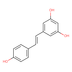
| HET Code: | STL |
|---|---|
| Formula: | C14H12O3 |
| Molecular weight: | 228.243 g/mol |
| DrugBank ID: | DB02709 |
| Buried Surface Area: | 35.32 % |
| Polar Surface area: | 60.69 Å2 |
| Number of | |
|---|---|
| H-Bond Acceptors: | 3 |
| H-Bond Donors: | 3 |
| Rings: | 2 |
| Aromatic rings: | 2 |
| Anionic atoms: | 0 |
| Cationic atoms: | 0 |
| Rule of Five Violation: | 0 |
| Rotatable Bonds: | 2 |
| X | Y | Z |
|---|---|---|
| 9.63735 | 12.8558 | -3.70165 |
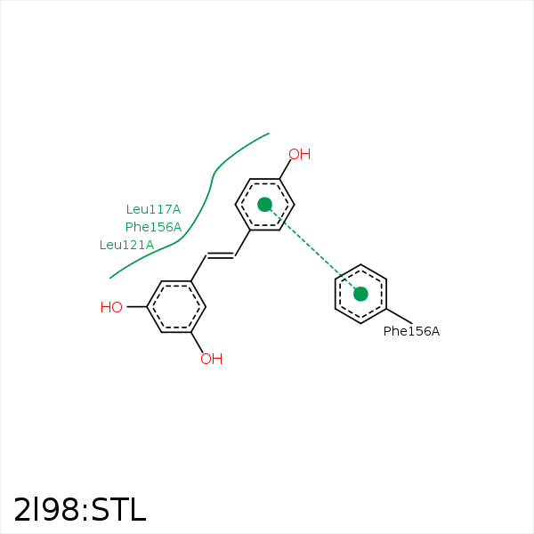


Represent the protein/ligand binding mode, centered on the ligand
Dashed lines represents hydrogen bonds and metal interactions
Green residue labels for amino acids with hydrophobic contacts (green lines) to the ligand
| Ligand | Protein | Interaction | |||
|---|---|---|---|---|---|
| Atom | Atom | Residue | Distance (Å) | Angle (°) | Type |
| C14 | CD2 | LEU- 100 | 4.36 | 0 | Hydrophobic |
| C12 | CD2 | LEU- 117 | 3.44 | 0 | Hydrophobic |
| C5 | CD1 | LEU- 121 | 3.82 | 0 | Hydrophobic |
| C10 | CD1 | LEU- 136 | 3.59 | 0 | Hydrophobic |
| C13 | CE1 | PHE- 153 | 3.28 | 0 | Hydrophobic |
| DuAr | DuAr | PHE- 156 | 3.84 | 0 | Aromatic Face/Face |
