sc-PDB
An Annotated Database of Druggable Binding Sites from the Protein DataBank
An Annotated Database of Druggable Binding Sites from the Protein DataBank
1.300 Å
X-ray
1997-08-13
| Name: | Streptavidin |
|---|---|
| ID: | SAV_STRAV |
| AC: | P22629 |
| Organism: | Streptomyces avidinii |
| Reign: | Bacteria |
| TaxID: | 1895 |
| EC Number: | / |
| Chain Name: | Percentage of Residues within binding site |
|---|---|
| A | 100 % |
| B-Factor: | 21.317 |
|---|---|
| Number of residues: | 23 |
| Including | |
| Standard Amino Acids: | 23 |
| Non Standard Amino Acids: | 0 |
| Water Molecules: | 0 |
| Cofactors: | |
| Metals: | |
| Ligandability | Volume (Å3) |
|---|---|
| 1.014 | 283.500 |
| % Hydrophobic | % Polar |
|---|---|
| 70.24 | 29.76 |
| According to VolSite | |
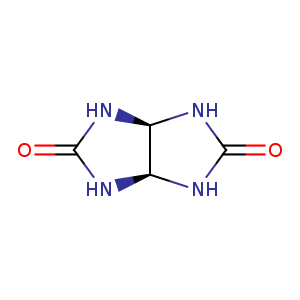
| HET Code: | GLL |
|---|---|
| Formula: | C4H6N4O2 |
| Molecular weight: | 142.116 g/mol |
| DrugBank ID: | DB03533 |
| Buried Surface Area: | 77.92 % |
| Polar Surface area: | 82.26 Å2 |
| Number of | |
|---|---|
| H-Bond Acceptors: | 2 |
| H-Bond Donors: | 4 |
| Rings: | 2 |
| Aromatic rings: | 0 |
| Anionic atoms: | 0 |
| Cationic atoms: | 0 |
| Rule of Five Violation: | 0 |
| Rotatable Bonds: | 0 |
| X | Y | Z |
|---|---|---|
| 11.9763 | 0.194 | -8.543 |
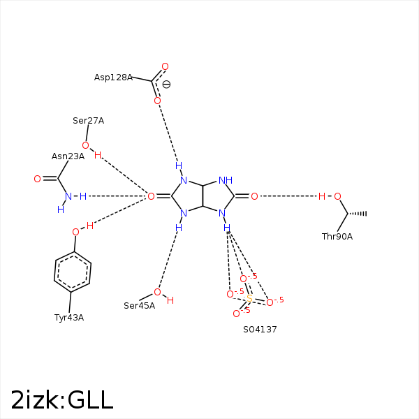


Represent the protein/ligand binding mode, centered on the ligand
Dashed lines represents hydrogen bonds and metal interactions
Green residue labels for amino acids with hydrophobic contacts (green lines) to the ligand
| Ligand | Protein | Interaction | |||
|---|---|---|---|---|---|
| Atom | Atom | Residue | Distance (Å) | Angle (°) | Type |
| O1 | ND2 | ASN- 23 | 2.9 | 144.35 | H-Bond (Protein Donor) |
| O1 | OG | SER- 27 | 2.78 | 143.87 | H-Bond (Protein Donor) |
| O1 | OH | TYR- 43 | 2.66 | 173.18 | H-Bond (Protein Donor) |
| N1 | OG | SER- 45 | 2.8 | 175.96 | H-Bond (Ligand Donor) |
| O1' | OG1 | THR- 90 | 2.66 | 169.91 | H-Bond (Protein Donor) |
| N2 | OD2 | ASP- 128 | 2.92 | 174.05 | H-Bond (Ligand Donor) |
