sc-PDB
An Annotated Database of Druggable Binding Sites from the Protein DataBank
An Annotated Database of Druggable Binding Sites from the Protein DataBank
1.500 Å
X-ray
2006-06-23
| Name: | Lipopolysaccharide core biosynthesis protein RfaG |
|---|---|
| ID: | RFAG_ECOLI |
| AC: | P25740 |
| Organism: | Escherichia coli |
| Reign: | Bacteria |
| TaxID: | 83333 |
| EC Number: | 2.4 |
| Chain Name: | Percentage of Residues within binding site |
|---|---|
| A | 100 % |
| B-Factor: | 8.075 |
|---|---|
| Number of residues: | 53 |
| Including | |
| Standard Amino Acids: | 47 |
| Non Standard Amino Acids: | 0 |
| Water Molecules: | 6 |
| Cofactors: | |
| Metals: | |
| Ligandability | Volume (Å3) |
|---|---|
| 0.221 | 874.125 |
| % Hydrophobic | % Polar |
|---|---|
| 32.82 | 67.18 |
| According to VolSite | |
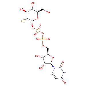
| HET Code: | U2F |
|---|---|
| Formula: | C15H21FN2O16P2 |
| Molecular weight: | 566.277 g/mol |
| DrugBank ID: | DB03488 |
| Buried Surface Area: | 77.53 % |
| Polar Surface area: | 296.59 Å2 |
| Number of | |
|---|---|
| H-Bond Acceptors: | 16 |
| H-Bond Donors: | 6 |
| Rings: | 3 |
| Aromatic rings: | 0 |
| Anionic atoms: | 2 |
| Cationic atoms: | 0 |
| Rule of Five Violation: | 3 |
| Rotatable Bonds: | 9 |
| X | Y | Z |
|---|---|---|
| -5.63092 | 10.8896 | -19.0942 |


Represent the protein/ligand binding mode, centered on the ligand
Dashed lines represents hydrogen bonds and metal interactions
Green residue labels for amino acids with hydrophobic contacts (green lines) to the ligand
| Ligand | Protein | Interaction | |||
|---|---|---|---|---|---|
| Atom | Atom | Residue | Distance (Å) | Angle (°) | Type |
| O1B | N | GLY- 15 | 2.75 | 168.49 | H-Bond (Protein Donor) |
| C1' | CD | ARG- 18 | 3.69 | 0 | Hydrophobic |
| C4' | CD | ARG- 18 | 3.98 | 0 | Hydrophobic |
| O6 | OD2 | ASP- 19 | 2.74 | 159.43 | H-Bond (Ligand Donor) |
| C6 | CE2 | PHE- 97 | 4.24 | 0 | Hydrophobic |
| C6 | CB | ALA- 99 | 3.87 | 0 | Hydrophobic |
| C4 | CD1 | LEU- 143 | 4.45 | 0 | Hydrophobic |
| C6 | CD1 | LEU- 143 | 3.82 | 0 | Hydrophobic |
| O2' | NH2 | ARG- 173 | 3.08 | 142.02 | H-Bond (Protein Donor) |
| C2' | CG1 | VAL- 202 | 4.18 | 0 | Hydrophobic |
| O2B | CZ | ARG- 208 | 3.83 | 0 | Ionic (Protein Cationic) |
| O2B | NH2 | ARG- 208 | 2.78 | 157.67 | H-Bond (Protein Donor) |
| O2B | NZ | LYS- 209 | 2.82 | 152.13 | H-Bond (Protein Donor) |
| O3A | NZ | LYS- 209 | 2.93 | 127.47 | H-Bond (Protein Donor) |
| O2B | NZ | LYS- 209 | 2.82 | 0 | Ionic (Protein Cationic) |
| N3 | O | ARG- 261 | 2.83 | 173.19 | H-Bond (Ligand Donor) |
| O7' | N | ARG- 261 | 2.98 | 165.35 | H-Bond (Protein Donor) |
| O3 | OE2 | GLU- 281 | 2.67 | 155.4 | H-Bond (Ligand Donor) |
| O3 | N | ALA- 283 | 2.96 | 166.11 | H-Bond (Protein Donor) |
| C4 | CB | ALA- 283 | 4.16 | 0 | Hydrophobic |
| O4 | N | GLY- 284 | 2.93 | 164.89 | H-Bond (Protein Donor) |
| O1A | N | ILE- 285 | 2.79 | 158.88 | H-Bond (Protein Donor) |
| C3' | CG2 | ILE- 285 | 4 | 0 | Hydrophobic |
| C6 | CD1 | ILE- 285 | 4.13 | 0 | Hydrophobic |
| O2A | N | VAL- 286 | 3.14 | 160.43 | H-Bond (Protein Donor) |
| C5' | CG1 | VAL- 286 | 4.46 | 0 | Hydrophobic |
| C2' | CG1 | VAL- 286 | 3.74 | 0 | Hydrophobic |
| O3' | OE2 | GLU- 289 | 2.63 | 174.35 | H-Bond (Ligand Donor) |
| O2' | OE2 | GLU- 289 | 3.09 | 131.14 | H-Bond (Ligand Donor) |
| O2' | OE1 | GLU- 289 | 2.65 | 162.74 | H-Bond (Ligand Donor) |
| O2A | O | HOH- 2583 | 2.73 | 138.17 | H-Bond (Protein Donor) |
| O1B | O | HOH- 2584 | 2.78 | 175.61 | H-Bond (Protein Donor) |
