sc-PDB
An Annotated Database of Druggable Binding Sites from the Protein DataBank
An Annotated Database of Druggable Binding Sites from the Protein DataBank
1.900 Å
X-ray
2006-10-02
| Name: | AGAP009194-PA |
|---|---|
| ID: | Q7PVS6_ANOGA |
| AC: | Q7PVS6 |
| Organism: | Anopheles gambiae |
| Reign: | Eukaryota |
| TaxID: | 7165 |
| EC Number: | / |
| Chain Name: | Percentage of Residues within binding site |
|---|---|
| A | 18 % |
| B | 82 % |
| B-Factor: | 21.205 |
|---|---|
| Number of residues: | 39 |
| Including | |
| Standard Amino Acids: | 39 |
| Non Standard Amino Acids: | 0 |
| Water Molecules: | 0 |
| Cofactors: | |
| Metals: | |
| Ligandability | Volume (Å3) |
|---|---|
| 1.032 | 843.750 |
| % Hydrophobic | % Polar |
|---|---|
| 65.60 | 34.40 |
| According to VolSite | |
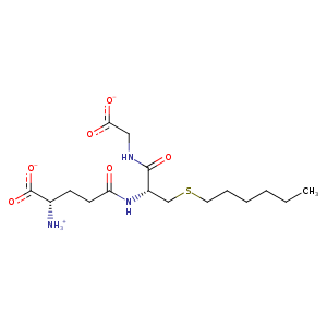
| HET Code: | GTX |
|---|---|
| Formula: | C16H28N3O6S |
| Molecular weight: | 390.475 g/mol |
| DrugBank ID: | - |
| Buried Surface Area: | 59.25 % |
| Polar Surface area: | 191.4 Å2 |
| Number of | |
|---|---|
| H-Bond Acceptors: | 7 |
| H-Bond Donors: | 3 |
| Rings: | 0 |
| Aromatic rings: | 0 |
| Anionic atoms: | 2 |
| Cationic atoms: | 1 |
| Rule of Five Violation: | 0 |
| Rotatable Bonds: | 15 |
| X | Y | Z |
|---|---|---|
| 4.08435 | 13.031 | 30.0052 |
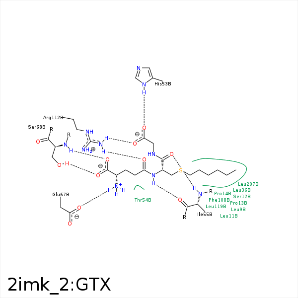


Represent the protein/ligand binding mode, centered on the ligand
Dashed lines represents hydrogen bonds and metal interactions
Green residue labels for amino acids with hydrophobic contacts (green lines) to the ligand
| Ligand | Protein | Interaction | |||
|---|---|---|---|---|---|
| Atom | Atom | Residue | Distance (Å) | Angle (°) | Type |
| C6S | CD2 | LEU- 9 | 4.05 | 0 | Hydrophobic |
| C6S | CD2 | LEU- 11 | 3.57 | 0 | Hydrophobic |
| SG2 | CB | SER- 12 | 4.27 | 0 | Hydrophobic |
| C4S | CB | SER- 12 | 4.14 | 0 | Hydrophobic |
| CG1 | CG | PRO- 14 | 3.92 | 0 | Hydrophobic |
| SG2 | CG | PRO- 14 | 3.83 | 0 | Hydrophobic |
| CB2 | CD1 | LEU- 36 | 3.68 | 0 | Hydrophobic |
| C3S | CD1 | LEU- 36 | 3.85 | 0 | Hydrophobic |
| O31 | ND1 | HIS- 53 | 3.09 | 137.5 | H-Bond (Protein Donor) |
| CG1 | CB | THR- 54 | 3.82 | 0 | Hydrophobic |
| N2 | O | ILE- 55 | 3.16 | 147.29 | H-Bond (Ligand Donor) |
| O2 | N | ILE- 55 | 2.96 | 161.07 | H-Bond (Protein Donor) |
| CB2 | CG1 | ILE- 55 | 4.18 | 0 | Hydrophobic |
| N1 | OE1 | GLU- 67 | 2.79 | 156.87 | H-Bond (Ligand Donor) |
| N1 | OE1 | GLU- 67 | 2.79 | 0 | Ionic (Ligand Cationic) |
| O11 | N | SER- 68 | 2.94 | 167.47 | H-Bond (Protein Donor) |
| O12 | OG | SER- 68 | 2.75 | 172.08 | H-Bond (Protein Donor) |
| CB1 | CE1 | PHE- 108 | 3.74 | 0 | Hydrophobic |
| CG1 | CZ | PHE- 108 | 4.29 | 0 | Hydrophobic |
| SG2 | CZ | PHE- 108 | 3.74 | 0 | Hydrophobic |
| OE1 | NH2 | ARG- 112 | 2.89 | 120.31 | H-Bond (Protein Donor) |
| O32 | NH1 | ARG- 112 | 3.38 | 147.1 | H-Bond (Protein Donor) |
| O32 | NH2 | ARG- 112 | 3.26 | 154.43 | H-Bond (Protein Donor) |
| O32 | CZ | ARG- 112 | 3.78 | 0 | Ionic (Protein Cationic) |
| C5S | CD1 | LEU- 119 | 3.91 | 0 | Hydrophobic |
| C5S | CD1 | LEU- 207 | 3.57 | 0 | Hydrophobic |
