sc-PDB
An Annotated Database of Druggable Binding Sites from the Protein DataBank
An Annotated Database of Druggable Binding Sites from the Protein DataBank
2.700 Å
X-ray
2006-05-30
| Name: | Biliverdin reductase A |
|---|---|
| ID: | BIEA_HUMAN |
| AC: | P53004 |
| Organism: | Homo sapiens |
| Reign: | Eukaryota |
| TaxID: | 9606 |
| EC Number: | 1.3.1.24 |
| Chain Name: | Percentage of Residues within binding site |
|---|---|
| C | 100 % |
| B-Factor: | 71.075 |
|---|---|
| Number of residues: | 32 |
| Including | |
| Standard Amino Acids: | 32 |
| Non Standard Amino Acids: | 0 |
| Water Molecules: | 0 |
| Cofactors: | |
| Metals: | |
| Ligandability | Volume (Å3) |
|---|---|
| 0.789 | 1144.125 |
| % Hydrophobic | % Polar |
|---|---|
| 42.77 | 57.23 |
| According to VolSite | |
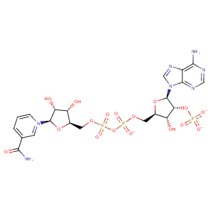
| HET Code: | NAP |
|---|---|
| Formula: | C21H25N7O17P3 |
| Molecular weight: | 740.381 g/mol |
| DrugBank ID: | DB03461 |
| Buried Surface Area: | 56.06 % |
| Polar Surface area: | 405.54 Å2 |
| Number of | |
|---|---|
| H-Bond Acceptors: | 21 |
| H-Bond Donors: | 5 |
| Rings: | 5 |
| Aromatic rings: | 3 |
| Anionic atoms: | 4 |
| Cationic atoms: | 1 |
| Rule of Five Violation: | 2 |
| Rotatable Bonds: | 13 |
| X | Y | Z |
|---|---|---|
| 7.95431 | 3.36287 | 23.9148 |
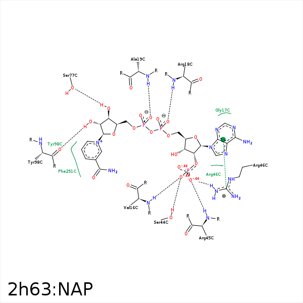


Represent the protein/ligand binding mode, centered on the ligand
Dashed lines represents hydrogen bonds and metal interactions
Green residue labels for amino acids with hydrophobic contacts (green lines) to the ligand
| Ligand | Protein | Interaction | |||
|---|---|---|---|---|---|
| Atom | Atom | Residue | Distance (Å) | Angle (°) | Type |
| O2X | N | VAL- 16 | 3.1 | 155.5 | H-Bond (Protein Donor) |
| O1A | NH1 | ARG- 18 | 3.49 | 136.41 | H-Bond (Protein Donor) |
| O2A | N | ARG- 18 | 2.67 | 171.8 | H-Bond (Protein Donor) |
| O2A | NH1 | ARG- 18 | 3.46 | 164.69 | H-Bond (Protein Donor) |
| O1N | CZ | ARG- 18 | 3.5 | 0 | Ionic (Protein Cationic) |
| O2N | N | ALA- 19 | 2.83 | 166.32 | H-Bond (Protein Donor) |
| C5D | CB | ALA- 19 | 4.28 | 0 | Hydrophobic |
| O2X | OG | SER- 44 | 2.67 | 160.7 | H-Bond (Protein Donor) |
| O3X | CZ | ARG- 45 | 3.05 | 0 | Ionic (Protein Cationic) |
| O3X | N | ARG- 45 | 2.67 | 165.14 | H-Bond (Protein Donor) |
| N1A | NH1 | ARG- 46 | 3.35 | 121.98 | H-Bond (Protein Donor) |
| O2X | NH2 | ARG- 46 | 2.99 | 144.7 | H-Bond (Protein Donor) |
| O2X | NE | ARG- 46 | 3.37 | 131.85 | H-Bond (Protein Donor) |
| O1X | CZ | ARG- 46 | 3.28 | 0 | Ionic (Protein Cationic) |
| O2X | CZ | ARG- 46 | 3.57 | 0 | Ionic (Protein Cationic) |
| DuAr | CZ | ARG- 46 | 3.64 | 16.41 | Pi/Cation |
| C4B | CG | GLU- 76 | 4.29 | 0 | Hydrophobic |
| O3D | OG | SER- 77 | 3.26 | 163.1 | H-Bond (Ligand Donor) |
| C4D | CB | GLU- 97 | 4.18 | 0 | Hydrophobic |
| O2D | O | TYR- 98 | 2.86 | 160.43 | H-Bond (Ligand Donor) |
| C5N | CE2 | TYR- 98 | 3.46 | 0 | Hydrophobic |
| C3D | CE1 | PHE- 162 | 4.44 | 0 | Hydrophobic |
| C2D | CD1 | PHE- 162 | 4.39 | 0 | Hydrophobic |
