sc-PDB
An Annotated Database of Druggable Binding Sites from the Protein DataBank
An Annotated Database of Druggable Binding Sites from the Protein DataBank
2.700 Å
X-ray
2006-04-09
| Name: | UDP-N-acetylglucosamine 4,6-dehydratase (inverting) |
|---|---|
| ID: | PSEB_HELPY |
| AC: | O25511 |
| Organism: | Helicobacter pylori |
| Reign: | Bacteria |
| TaxID: | 85962 |
| EC Number: | 4.2.1.115 |
| Chain Name: | Percentage of Residues within binding site |
|---|---|
| A | 98 % |
| B | 2 % |
| B-Factor: | 44.462 |
|---|---|
| Number of residues: | 43 |
| Including | |
| Standard Amino Acids: | 41 |
| Non Standard Amino Acids: | 1 |
| Water Molecules: | 1 |
| Cofactors: | NAP |
| Metals: | |
| Ligandability | Volume (Å3) |
|---|---|
| 0.748 | 1154.250 |
| % Hydrophobic | % Polar |
|---|---|
| 40.94 | 59.06 |
| According to VolSite | |
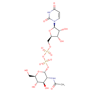
| HET Code: | UD1 |
|---|---|
| Formula: | C17H25N3O17P2 |
| Molecular weight: | 605.338 g/mol |
| DrugBank ID: | DB03397 |
| Buried Surface Area: | 67.3 % |
| Polar Surface area: | 325.69 Å2 |
| Number of | |
|---|---|
| H-Bond Acceptors: | 17 |
| H-Bond Donors: | 7 |
| Rings: | 3 |
| Aromatic rings: | 0 |
| Anionic atoms: | 2 |
| Cationic atoms: | 0 |
| Rule of Five Violation: | 3 |
| Rotatable Bonds: | 10 |
| X | Y | Z |
|---|---|---|
| 10.0194 | 27.7095 | 4.16195 |
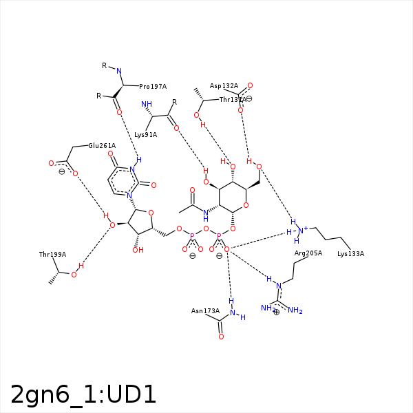


Represent the protein/ligand binding mode, centered on the ligand
Dashed lines represents hydrogen bonds and metal interactions
Green residue labels for amino acids with hydrophobic contacts (green lines) to the ligand
| Ligand | Protein | Interaction | |||
|---|---|---|---|---|---|
| Atom | Atom | Residue | Distance (Å) | Angle (°) | Type |
| C8' | CG | LYS- 91 | 3.76 | 0 | Hydrophobic |
| O3' | O | LYS- 91 | 3.36 | 172.26 | H-Bond (Ligand Donor) |
| C8' | CB | HIS- 92 | 4.37 | 0 | Hydrophobic |
| O4' | OG1 | THR- 131 | 3.1 | 164.88 | H-Bond (Protein Donor) |
| O6' | OD1 | ASP- 132 | 3.38 | 155.32 | H-Bond (Ligand Donor) |
| O6' | OD2 | ASP- 132 | 3.15 | 146.64 | H-Bond (Ligand Donor) |
| O1' | NZ | LYS- 133 | 3.5 | 125.84 | H-Bond (Protein Donor) |
| O6' | NZ | LYS- 133 | 2.82 | 130.89 | H-Bond (Protein Donor) |
| O2B | NZ | LYS- 133 | 3.08 | 173.09 | H-Bond (Protein Donor) |
| O2B | NZ | LYS- 133 | 3.08 | 0 | Ionic (Protein Cationic) |
| C5' | CD | LYS- 133 | 3.85 | 0 | Hydrophobic |
| C6' | CB | ASN- 173 | 4.36 | 0 | Hydrophobic |
| O2B | ND2 | ASN- 173 | 3.05 | 126.53 | H-Bond (Protein Donor) |
| N3 | O | PRO- 197 | 3.09 | 144.04 | H-Bond (Ligand Donor) |
| O2' | OG1 | THR- 199 | 3 | 153.86 | H-Bond (Protein Donor) |
| C3B | CG | MET- 203 | 4.09 | 0 | Hydrophobic |
| O2B | NE | ARG- 205 | 3.08 | 168.29 | H-Bond (Protein Donor) |
| C4B | CG | ARG- 205 | 4.29 | 0 | Hydrophobic |
| C1B | CE | MET- 239 | 3.46 | 0 | Hydrophobic |
| C4B | SD | MET- 239 | 3.58 | 0 | Hydrophobic |
| C3B | CG | MET- 239 | 4.16 | 0 | Hydrophobic |
| O1A | CZ | ARG- 258 | 2.97 | 0 | Ionic (Protein Cationic) |
| O2' | OE2 | GLU- 261 | 2.8 | 158.6 | H-Bond (Ligand Donor) |
| C4' | C3N | NAP- 334 | 3.86 | 0 | Hydrophobic |
| C6' | C4N | NAP- 334 | 3.88 | 0 | Hydrophobic |
