sc-PDB
An Annotated Database of Druggable Binding Sites from the Protein DataBank
An Annotated Database of Druggable Binding Sites from the Protein DataBank
2.200 Å
X-ray
2006-01-05
| Name: | Urocanate hydratase |
|---|---|
| ID: | HUTU_BACSU |
| AC: | P25503 |
| Organism: | Bacillus subtilis |
| Reign: | Bacteria |
| TaxID: | 224308 |
| EC Number: | 4.2.1.49 |
| Chain Name: | Percentage of Residues within binding site |
|---|---|
| C | 100 % |
| B-Factor: | 21.701 |
|---|---|
| Number of residues: | 65 |
| Including | |
| Standard Amino Acids: | 63 |
| Non Standard Amino Acids: | 0 |
| Water Molecules: | 2 |
| Cofactors: | |
| Metals: | |
| Ligandability | Volume (Å3) |
|---|---|
| 0.123 | 374.625 |
| % Hydrophobic | % Polar |
|---|---|
| 51.35 | 48.65 |
| According to VolSite | |
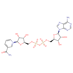
| HET Code: | NAD |
|---|---|
| Formula: | C21H26N7O14P2 |
| Molecular weight: | 662.417 g/mol |
| DrugBank ID: | - |
| Buried Surface Area: | 83.36 % |
| Polar Surface area: | 343.54 Å2 |
| Number of | |
|---|---|
| H-Bond Acceptors: | 18 |
| H-Bond Donors: | 6 |
| Rings: | 5 |
| Aromatic rings: | 3 |
| Anionic atoms: | 2 |
| Cationic atoms: | 1 |
| Rule of Five Violation: | 3 |
| Rotatable Bonds: | 11 |
| X | Y | Z |
|---|---|---|
| 42.5618 | 28.8446 | 0.977682 |


Represent the protein/ligand binding mode, centered on the ligand
Dashed lines represents hydrogen bonds and metal interactions
Green residue labels for amino acids with hydrophobic contacts (green lines) to the ligand
| Ligand | Protein | Interaction | |||
|---|---|---|---|---|---|
| Atom | Atom | Residue | Distance (Å) | Angle (°) | Type |
| O2N | N | GLY- 49 | 2.86 | 139.93 | H-Bond (Protein Donor) |
| O2A | N | GLY- 50 | 2.73 | 147.71 | H-Bond (Protein Donor) |
| O2D | NE2 | GLN- 127 | 3.24 | 143.99 | H-Bond (Protein Donor) |
| C4N | CD1 | ILE- 141 | 3.38 | 0 | Hydrophobic |
| O1A | N | GLY- 173 | 3.31 | 153.42 | H-Bond (Protein Donor) |
| O1N | N | MET- 174 | 3.07 | 129.39 | H-Bond (Protein Donor) |
| O2N | N | MET- 174 | 3.16 | 158.22 | H-Bond (Protein Donor) |
| C5N | CG | MET- 174 | 3.56 | 0 | Hydrophobic |
| O1N | N | GLY- 175 | 2.79 | 174.06 | H-Bond (Protein Donor) |
| O3B | OE1 | GLU- 193 | 2.85 | 170.61 | H-Bond (Ligand Donor) |
| O3B | OE2 | GLU- 193 | 3.16 | 125.16 | H-Bond (Ligand Donor) |
| O2B | OE2 | GLU- 193 | 2.61 | 168.73 | H-Bond (Ligand Donor) |
| N3A | N | VAL- 194 | 3.42 | 156.5 | H-Bond (Protein Donor) |
| O1A | NH1 | ARG- 198 | 3.12 | 123.19 | H-Bond (Protein Donor) |
| O1A | CZ | ARG- 198 | 3.88 | 0 | Ionic (Protein Cationic) |
| C3B | CG | ARG- 198 | 4.26 | 0 | Hydrophobic |
| N6A | OD1 | ASN- 239 | 2.91 | 145.14 | H-Bond (Ligand Donor) |
| N1A | N | ALA- 240 | 3.01 | 174.92 | H-Bond (Protein Donor) |
| O4D | NE2 | GLN- 260 | 3.05 | 170.12 | H-Bond (Protein Donor) |
| C4D | CG | GLN- 260 | 4.13 | 0 | Hydrophobic |
| O2A | OG | SER- 262 | 2.91 | 165.46 | H-Bond (Protein Donor) |
| C5B | CB | SER- 262 | 3.93 | 0 | Hydrophobic |
| C3D | CB | SER- 262 | 4.33 | 0 | Hydrophobic |
| O3D | ND1 | HIS- 264 | 2.8 | 167.86 | H-Bond (Ligand Donor) |
| N7A | N | VAL- 271 | 3.01 | 167.32 | H-Bond (Protein Donor) |
| N6A | O | VAL- 271 | 3.46 | 134.35 | H-Bond (Ligand Donor) |
| N7N | O | TYR- 319 | 3.14 | 135.55 | H-Bond (Ligand Donor) |
| C4D | CB | ASN- 321 | 4.04 | 0 | Hydrophobic |
| O2D | O | GLY- 489 | 2.82 | 149.89 | H-Bond (Ligand Donor) |
| O1N | O | HOH- 7602 | 2.72 | 179.93 | H-Bond (Protein Donor) |
