sc-PDB
An Annotated Database of Druggable Binding Sites from the Protein DataBank
An Annotated Database of Druggable Binding Sites from the Protein DataBank
1.800 Å
X-ray
2006-01-02
| Name: | Adenylylsulfate reductase, subunit A (AprA) |
|---|---|
| ID: | O28603_ARCFU |
| AC: | O28603 |
| Organism: | Archaeoglobus fulgidus |
| Reign: | Archaea |
| TaxID: | 224325 |
| EC Number: | / |
| Chain Name: | Percentage of Residues within binding site |
|---|---|
| C | 97 % |
| D | 3 % |
| B-Factor: | 8.920 |
|---|---|
| Number of residues: | 83 |
| Including | |
| Standard Amino Acids: | 73 |
| Non Standard Amino Acids: | 0 |
| Water Molecules: | 10 |
| Cofactors: | |
| Metals: | |
| Ligandability | Volume (Å3) |
|---|---|
| 0.892 | 776.250 |
| % Hydrophobic | % Polar |
|---|---|
| 48.70 | 51.30 |
| According to VolSite | |
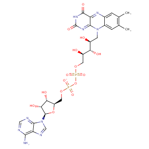
| HET Code: | FAD |
|---|---|
| Formula: | C27H31N9O15P2 |
| Molecular weight: | 783.534 g/mol |
| DrugBank ID: | DB03147 |
| Buried Surface Area: | 82.43 % |
| Polar Surface area: | 381.7 Å2 |
| Number of | |
|---|---|
| H-Bond Acceptors: | 22 |
| H-Bond Donors: | 7 |
| Rings: | 6 |
| Aromatic rings: | 3 |
| Anionic atoms: | 2 |
| Cationic atoms: | 0 |
| Rule of Five Violation: | 3 |
| Rotatable Bonds: | 13 |
| X | Y | Z |
|---|---|---|
| 42.72 | -0.592792 | 82.7543 |


Represent the protein/ligand binding mode, centered on the ligand
Dashed lines represents hydrogen bonds and metal interactions
Green residue labels for amino acids with hydrophobic contacts (green lines) to the ligand
| Ligand | Protein | Interaction | |||
|---|---|---|---|---|---|
| Atom | Atom | Residue | Distance (Å) | Angle (°) | Type |
| O1A | N | PHE- 2032 | 3.14 | 169.3 | H-Bond (Protein Donor) |
| C4' | CB | PHE- 2032 | 4.23 | 0 | Hydrophobic |
| O1P | N | SER- 2033 | 2.96 | 159.81 | H-Bond (Protein Donor) |
| O1P | OG | SER- 2033 | 2.66 | 159.57 | H-Bond (Protein Donor) |
| O3B | OE2 | GLU- 2056 | 2.64 | 155.7 | H-Bond (Ligand Donor) |
| O3B | OE1 | GLU- 2056 | 3.1 | 122.68 | H-Bond (Ligand Donor) |
| O2B | OE1 | GLU- 2056 | 2.51 | 158.39 | H-Bond (Ligand Donor) |
| C1B | CG | LYS- 2057 | 4.47 | 0 | Hydrophobic |
| N3A | N | LYS- 2057 | 3.17 | 140.48 | H-Bond (Protein Donor) |
| C3B | CB | SER- 2063 | 4.18 | 0 | Hydrophobic |
| O3B | OG | SER- 2063 | 2.77 | 143.35 | H-Bond (Protein Donor) |
| O2A | N | ALA- 2065 | 2.96 | 165.85 | H-Bond (Protein Donor) |
| C2' | CB | ALA- 2065 | 4.45 | 0 | Hydrophobic |
| C8M | CB | ALA- 2065 | 3.56 | 0 | Hydrophobic |
| C6 | CD2 | LEU- 2070 | 4.26 | 0 | Hydrophobic |
| C9A | CD2 | LEU- 2070 | 4.02 | 0 | Hydrophobic |
| C2' | CD1 | LEU- 2070 | 4.37 | 0 | Hydrophobic |
| N3 | O | ALA- 2072 | 2.58 | 134.92 | H-Bond (Ligand Donor) |
| O2 | N | ASN- 2074 | 2.92 | 178.2 | H-Bond (Protein Donor) |
| N6A | O | ILE- 2176 | 2.83 | 160.72 | H-Bond (Ligand Donor) |
| N1A | N | ILE- 2176 | 3.07 | 167.3 | H-Bond (Protein Donor) |
| C7M | CE2 | TRP- 2234 | 3.88 | 0 | Hydrophobic |
| C8M | CD1 | TYR- 2235 | 3.55 | 0 | Hydrophobic |
| C8M | CB | ALA- 2236 | 4.25 | 0 | Hydrophobic |
| O2A | OD2 | ASP- 2239 | 2.7 | 172.63 | H-Bond (Protein Donor) |
| N6A | OG | SER- 2242 | 2.95 | 163.46 | H-Bond (Ligand Donor) |
| C6 | CE | MET- 2365 | 3.36 | 0 | Hydrophobic |
| C3' | CB | SER- 2397 | 4.49 | 0 | Hydrophobic |
| C5' | CB | SER- 2397 | 4.11 | 0 | Hydrophobic |
| O3' | OG | SER- 2397 | 2.75 | 165.48 | H-Bond (Ligand Donor) |
| O2P | N | ASP- 2439 | 2.81 | 175.95 | H-Bond (Protein Donor) |
| C1' | CD2 | PHE- 2448 | 4.39 | 0 | Hydrophobic |
| N1 | N | SER- 2449 | 3.22 | 151.53 | H-Bond (Protein Donor) |
| O2 | N | SER- 2449 | 3.08 | 147.3 | H-Bond (Protein Donor) |
| C5' | CB | SER- 2452 | 4.02 | 0 | Hydrophobic |
| C7M | CZ2 | TRP- 2748 | 4.32 | 0 | Hydrophobic |
| C8M | CH2 | TRP- 2748 | 3.77 | 0 | Hydrophobic |
| O1P | O | HOH- 5008 | 2.76 | 167.68 | H-Bond (Protein Donor) |
| O1A | O | HOH- 5048 | 2.9 | 179.94 | H-Bond (Protein Donor) |
| O2B | O | HOH- 5059 | 2.85 | 179.97 | H-Bond (Protein Donor) |
| O2P | O | HOH- 5323 | 2.71 | 171.87 | H-Bond (Protein Donor) |
