sc-PDB
An Annotated Database of Druggable Binding Sites from the Protein DataBank
An Annotated Database of Druggable Binding Sites from the Protein DataBank
2.500 Å
X-ray
2005-11-22
| Name: | Guanylate kinase |
|---|---|
| ID: | KGUA_ECOLI |
| AC: | P60546 |
| Organism: | Escherichia coli |
| Reign: | Bacteria |
| TaxID: | 83333 |
| EC Number: | 2.7.4.8 |
| Chain Name: | Percentage of Residues within binding site |
|---|---|
| B | 100 % |
| B-Factor: | 48.951 |
|---|---|
| Number of residues: | 49 |
| Including | |
| Standard Amino Acids: | 47 |
| Non Standard Amino Acids: | 0 |
| Water Molecules: | 2 |
| Cofactors: | |
| Metals: | |
| Ligandability | Volume (Å3) |
|---|---|
| 0.139 | 759.375 |
| % Hydrophobic | % Polar |
|---|---|
| 28.00 | 72.00 |
| According to VolSite | |
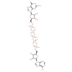
| HET Code: | G5P |
|---|---|
| Formula: | C20H24N10O23P5 |
| Molecular weight: | 927.327 g/mol |
| DrugBank ID: | - |
| Buried Surface Area: | 51.84 % |
| Polar Surface area: | 559.37 Å2 |
| Number of | |
|---|---|
| H-Bond Acceptors: | 30 |
| H-Bond Donors: | 7 |
| Rings: | 6 |
| Aromatic rings: | 3 |
| Anionic atoms: | 5 |
| Cationic atoms: | 0 |
| Rule of Five Violation: | 3 |
| Rotatable Bonds: | 16 |
| X | Y | Z |
|---|---|---|
| 7.63843 | 17.3363 | 39.4905 |


Represent the protein/ligand binding mode, centered on the ligand
Dashed lines represents hydrogen bonds and metal interactions
Green residue labels for amino acids with hydrophobic contacts (green lines) to the ligand
| Ligand | Protein | Interaction | |||
|---|---|---|---|---|---|
| Atom | Atom | Residue | Distance (Å) | Angle (°) | Type |
| O2G | OG | SER- 13 | 3.34 | 155.98 | H-Bond (Protein Donor) |
| O2D | N | GLY- 14 | 3.12 | 133.79 | H-Bond (Protein Donor) |
| O2D | N | GLY- 16 | 3.13 | 142.15 | H-Bond (Protein Donor) |
| O1D | NZ | LYS- 17 | 3.4 | 178.05 | H-Bond (Protein Donor) |
| O1D | NZ | LYS- 17 | 3.4 | 0 | Ionic (Protein Cationic) |
| O1B | NZ | LYS- 17 | 3.35 | 0 | Ionic (Protein Cationic) |
| O1D | N | SER- 18 | 2.76 | 166.58 | H-Bond (Protein Donor) |
| O1D | OG | SER- 18 | 3.47 | 156.41 | H-Bond (Protein Donor) |
| O3D | OG | SER- 18 | 2.77 | 134.04 | H-Bond (Protein Donor) |
| O1E | N | SER- 19 | 3.38 | 142.97 | H-Bond (Protein Donor) |
| O2E | OG | SER- 19 | 2.63 | 159.53 | H-Bond (Protein Donor) |
| O6 | OG | SER- 38 | 2.65 | 148.66 | H-Bond (Protein Donor) |
| O1A | CZ | ARG- 42 | 3.72 | 0 | Ionic (Protein Cationic) |
| O2A | CZ | ARG- 42 | 3.55 | 0 | Ionic (Protein Cationic) |
| O1A | NH2 | ARG- 42 | 3.12 | 155.56 | H-Bond (Protein Donor) |
| O1A | NE | ARG- 42 | 3.43 | 141.77 | H-Bond (Protein Donor) |
| O2A | CZ | ARG- 45 | 3.45 | 0 | Ionic (Protein Cationic) |
| O1A | OH | TYR- 54 | 2.7 | 138.32 | H-Bond (Protein Donor) |
| N1 | OE1 | GLU- 73 | 3.08 | 143.98 | H-Bond (Ligand Donor) |
| N1 | OE2 | GLU- 73 | 3.15 | 145.41 | H-Bond (Ligand Donor) |
| N2 | OE1 | GLU- 73 | 2.97 | 148.7 | H-Bond (Ligand Donor) |
| C4' | CG1 | VAL- 77 | 4.17 | 0 | Hydrophobic |
| C1' | CG2 | VAL- 77 | 4.21 | 0 | Hydrophobic |
| N2 | OD1 | ASP- 104 | 2.77 | 160.72 | H-Bond (Ligand Donor) |
| N3 | N | ASP- 104 | 3.17 | 165.15 | H-Bond (Protein Donor) |
| C1' | CB | ASP- 104 | 4.17 | 0 | Hydrophobic |
| O2F | O | ARG- 134 | 2.81 | 161.97 | H-Bond (Ligand Donor) |
| O1D | O | HOH- 609 | 2.76 | 142.04 | H-Bond (Protein Donor) |
| O2' | O | HOH- 610 | 2.81 | 179.96 | H-Bond (Protein Donor) |
