sc-PDB
An Annotated Database of Druggable Binding Sites from the Protein DataBank
An Annotated Database of Druggable Binding Sites from the Protein DataBank
2.100 Å
X-ray
2004-12-23
| Name: | NADPH:ferredoxin reductase |
|---|---|
| ID: | Q9L6V3_RHOCA |
| AC: | Q9L6V3 |
| Organism: | Rhodobacter capsulatus |
| Reign: | Bacteria |
| TaxID: | 1061 |
| EC Number: | / |
| Chain Name: | Percentage of Residues within binding site |
|---|---|
| C | 100 % |
| B-Factor: | 38.105 |
|---|---|
| Number of residues: | 43 |
| Including | |
| Standard Amino Acids: | 40 |
| Non Standard Amino Acids: | 0 |
| Water Molecules: | 3 |
| Cofactors: | |
| Metals: | |
| Ligandability | Volume (Å3) |
|---|---|
| 0.960 | 1042.875 |
| % Hydrophobic | % Polar |
|---|---|
| 46.93 | 53.07 |
| According to VolSite | |
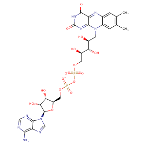
| HET Code: | FAD |
|---|---|
| Formula: | C27H31N9O15P2 |
| Molecular weight: | 783.534 g/mol |
| DrugBank ID: | DB03147 |
| Buried Surface Area: | 64.11 % |
| Polar Surface area: | 381.7 Å2 |
| Number of | |
|---|---|
| H-Bond Acceptors: | 22 |
| H-Bond Donors: | 7 |
| Rings: | 6 |
| Aromatic rings: | 3 |
| Anionic atoms: | 2 |
| Cationic atoms: | 0 |
| Rule of Five Violation: | 3 |
| Rotatable Bonds: | 13 |
| X | Y | Z |
|---|---|---|
| 32.1149 | 28.4407 | -4.31374 |
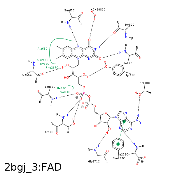


Represent the protein/ligand binding mode, centered on the ligand
Dashed lines represents hydrogen bonds and metal interactions
Green residue labels for amino acids with hydrophobic contacts (green lines) to the ligand
| Ligand | Protein | Interaction | |||
|---|---|---|---|---|---|
| Atom | Atom | Residue | Distance (Å) | Angle (°) | Type |
| C7M | CD1 | PHE- 49 | 3.87 | 0 | Hydrophobic |
| O1A | NE | ARG- 64 | 3.38 | 149.47 | H-Bond (Protein Donor) |
| O1P | NH2 | ARG- 64 | 3.26 | 121.1 | H-Bond (Protein Donor) |
| O1P | NE | ARG- 64 | 3.15 | 124.26 | H-Bond (Protein Donor) |
| C2' | CB | ARG- 64 | 4.08 | 0 | Hydrophobic |
| C3' | CG | ARG- 64 | 3.87 | 0 | Hydrophobic |
| O1P | CZ | ARG- 64 | 3.54 | 0 | Ionic (Protein Cationic) |
| O2' | O | ALA- 65 | 2.73 | 167.47 | H-Bond (Ligand Donor) |
| C7 | CB | ALA- 65 | 3.57 | 0 | Hydrophobic |
| C8 | CB | ALA- 65 | 3.56 | 0 | Hydrophobic |
| C8 | CB | ALA- 65 | 3.56 | 0 | Hydrophobic |
| C2' | CE1 | TYR- 66 | 4.22 | 0 | Hydrophobic |
| C4' | CE1 | TYR- 66 | 4.19 | 0 | Hydrophobic |
| O4' | OH | TYR- 66 | 2.76 | 138.84 | H-Bond (Ligand Donor) |
| O4 | N | SER- 67 | 3.17 | 131 | H-Bond (Protein Donor) |
| N5 | N | SER- 67 | 3.27 | 151.69 | H-Bond (Protein Donor) |
| N3 | O | TYR- 80 | 2.81 | 169.6 | H-Bond (Ligand Donor) |
| O2 | N | ILE- 82 | 2.98 | 167.74 | H-Bond (Protein Donor) |
| C5' | CG2 | ILE- 82 | 4.04 | 0 | Hydrophobic |
| C5B | CG1 | VAL- 84 | 4.14 | 0 | Hydrophobic |
| C1B | CG2 | VAL- 84 | 4.4 | 0 | Hydrophobic |
| O1P | N | LEU- 89 | 2.96 | 175.16 | H-Bond (Protein Donor) |
| O2P | N | LEU- 89 | 3.43 | 120.35 | H-Bond (Protein Donor) |
| O2P | OG1 | THR- 90 | 2.79 | 178.71 | H-Bond (Protein Donor) |
| O2P | N | THR- 90 | 2.88 | 169.46 | H-Bond (Protein Donor) |
| C5' | CG2 | THR- 90 | 3.63 | 0 | Hydrophobic |
| N6A | OG1 | THR- 130 | 2.73 | 159.28 | H-Bond (Ligand Donor) |
| N1A | OG1 | THR- 130 | 3.47 | 120.44 | H-Bond (Protein Donor) |
| C7M | CG | GLU- 264 | 3.96 | 0 | Hydrophobic |
| C1' | CB | PHE- 267 | 3.86 | 0 | Hydrophobic |
| C2B | CD1 | PHE- 267 | 3.97 | 0 | Hydrophobic |
| DuAr | DuAr | PHE- 267 | 3.78 | 0 | Aromatic Face/Face |
| C8M | CG2 | VAL- 268 | 4.3 | 0 | Hydrophobic |
| O2B | N | GLY- 271 | 2.91 | 129.26 | H-Bond (Protein Donor) |
| N3A | N | ILE- 272 | 3.22 | 167.96 | H-Bond (Protein Donor) |
| O4 | O | HOH- 2080 | 2.7 | 179.99 | H-Bond (Protein Donor) |
