sc-PDB
An Annotated Database of Druggable Binding Sites from the Protein DataBank
An Annotated Database of Druggable Binding Sites from the Protein DataBank
1.500 Å
X-ray
2005-08-16
| Name: | Alpha-1,3-mannosyl-glycoprotein 2-beta-N-acetylglucosaminyltransferase |
|---|---|
| ID: | MGAT1_RABIT |
| AC: | P27115 |
| Organism: | Oryctolagus cuniculus |
| Reign: | Eukaryota |
| TaxID: | 9986 |
| EC Number: | 2.4.1.101 |
| Chain Name: | Percentage of Residues within binding site |
|---|---|
| A | 100 % |
| B-Factor: | 13.012 |
|---|---|
| Number of residues: | 51 |
| Including | |
| Standard Amino Acids: | 42 |
| Non Standard Amino Acids: | 1 |
| Water Molecules: | 8 |
| Cofactors: | |
| Metals: | MN |
| Ligandability | Volume (Å3) |
|---|---|
| 0.077 | 455.625 |
| % Hydrophobic | % Polar |
|---|---|
| 41.48 | 58.52 |
| According to VolSite | |
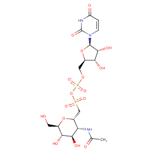
| HET Code: | UDM |
|---|---|
| Formula: | C18H27N3O16P2 |
| Molecular weight: | 603.365 g/mol |
| DrugBank ID: | - |
| Buried Surface Area: | 64.65 % |
| Polar Surface area: | 316.45 Å2 |
| Number of | |
|---|---|
| H-Bond Acceptors: | 16 |
| H-Bond Donors: | 7 |
| Rings: | 3 |
| Aromatic rings: | 0 |
| Anionic atoms: | 2 |
| Cationic atoms: | 0 |
| Rule of Five Violation: | 3 |
| Rotatable Bonds: | 10 |
| X | Y | Z |
|---|---|---|
| 1.42862 | 19.1009 | 11.1656 |
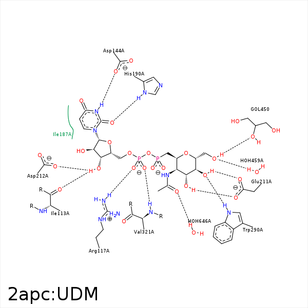


Represent the protein/ligand binding mode, centered on the ligand
Dashed lines represents hydrogen bonds and metal interactions
Green residue labels for amino acids with hydrophobic contacts (green lines) to the ligand
| Ligand | Protein | Interaction | |||
|---|---|---|---|---|---|
| Atom | Atom | Residue | Distance (Å) | Angle (°) | Type |
| O3D | O | ILE- 113 | 2.87 | 128.8 | H-Bond (Ligand Donor) |
| C3D | CG2 | ILE- 113 | 3.91 | 0 | Hydrophobic |
| C2D | CB | CYS- 115 | 4.27 | 0 | Hydrophobic |
| O1A | NH2 | ARG- 117 | 2.84 | 148.82 | H-Bond (Protein Donor) |
| O1A | NH1 | ARG- 117 | 3.41 | 127.85 | H-Bond (Protein Donor) |
| O1A | CZ | ARG- 117 | 3.54 | 0 | Ionic (Protein Cationic) |
| O2A | CZ | ARG- 117 | 3.83 | 0 | Ionic (Protein Cationic) |
| N3 | OD2 | ASP- 144 | 2.86 | 166.91 | H-Bond (Ligand Donor) |
| CB | CE2 | TYR- 184 | 4.46 | 0 | Hydrophobic |
| C5' | CD1 | ILE- 187 | 4.13 | 0 | Hydrophobic |
| C6' | CG2 | ILE- 187 | 4.11 | 0 | Hydrophobic |
| CB | CD1 | ILE- 187 | 4.36 | 0 | Hydrophobic |
| C1D | CG1 | ILE- 187 | 4.17 | 0 | Hydrophobic |
| C5D | CD1 | ILE- 187 | 3.88 | 0 | Hydrophobic |
| O2 | ND1 | HIS- 190 | 2.72 | 155.34 | H-Bond (Protein Donor) |
| O3' | OE2 | GLU- 211 | 2.63 | 166.1 | H-Bond (Ligand Donor) |
| O4' | OE1 | GLU- 211 | 2.67 | 164.97 | H-Bond (Ligand Donor) |
| C4D | CG | GLU- 211 | 4.04 | 0 | Hydrophobic |
| O3D | OD1 | ASP- 212 | 3.08 | 133.51 | H-Bond (Ligand Donor) |
| C8' | CD2 | LEU- 269 | 3.84 | 0 | Hydrophobic |
| C4' | CZ2 | TRP- 290 | 4.11 | 0 | Hydrophobic |
| C6' | CE2 | TRP- 290 | 4.23 | 0 | Hydrophobic |
| O4' | NE1 | TRP- 290 | 2.91 | 134.04 | H-Bond (Protein Donor) |
| O2A | N | VAL- 321 | 2.88 | 154.64 | H-Bond (Protein Donor) |
| C8' | CD1 | LEU- 331 | 3.56 | 0 | Hydrophobic |
| O1A | MN | MN- 448 | 2.24 | 0 | Metal Acceptor |
| O1B | MN | MN- 448 | 2.17 | 0 | Metal Acceptor |
| O6' | O | HOH- 459 | 2.84 | 179.97 | H-Bond (Protein Donor) |
| O7' | O | HOH- 646 | 2.89 | 179.95 | H-Bond (Protein Donor) |
