sc-PDB
An Annotated Database of Druggable Binding Sites from the Protein DataBank
An Annotated Database of Druggable Binding Sites from the Protein DataBank
1.840 Å
X-ray
2005-07-26
| Name: | 3-hydroxybutyrate dehydrogenase type 2 |
|---|---|
| ID: | BDH2_HUMAN |
| AC: | Q9BUT1 |
| Organism: | Homo sapiens |
| Reign: | Eukaryota |
| TaxID: | 9606 |
| EC Number: | / |
| Chain Name: | Percentage of Residues within binding site |
|---|---|
| B | 100 % |
| B-Factor: | 28.381 |
|---|---|
| Number of residues: | 50 |
| Including | |
| Standard Amino Acids: | 47 |
| Non Standard Amino Acids: | 0 |
| Water Molecules: | 3 |
| Cofactors: | |
| Metals: | |
| Ligandability | Volume (Å3) |
|---|---|
| 1.015 | 982.125 |
| % Hydrophobic | % Polar |
|---|---|
| 46.05 | 53.95 |
| According to VolSite | |
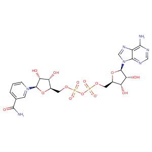
| HET Code: | NAD |
|---|---|
| Formula: | C21H26N7O14P2 |
| Molecular weight: | 662.417 g/mol |
| DrugBank ID: | - |
| Buried Surface Area: | 71.15 % |
| Polar Surface area: | 343.54 Å2 |
| Number of | |
|---|---|
| H-Bond Acceptors: | 18 |
| H-Bond Donors: | 6 |
| Rings: | 5 |
| Aromatic rings: | 3 |
| Anionic atoms: | 2 |
| Cationic atoms: | 1 |
| Rule of Five Violation: | 3 |
| Rotatable Bonds: | 11 |
| X | Y | Z |
|---|---|---|
| 39.5411 | 4.50348 | 52.0458 |


Represent the protein/ligand binding mode, centered on the ligand
Dashed lines represents hydrogen bonds and metal interactions
Green residue labels for amino acids with hydrophobic contacts (green lines) to the ligand
| Ligand | Protein | Interaction | |||
|---|---|---|---|---|---|
| Atom | Atom | Residue | Distance (Å) | Angle (°) | Type |
| C4B | CB | ALA- 13 | 3.87 | 0 | Hydrophobic |
| C1B | CB | ALA- 13 | 4.03 | 0 | Hydrophobic |
| O1A | NE2 | GLN- 16 | 2.87 | 166.66 | H-Bond (Protein Donor) |
| O5B | NE2 | GLN- 16 | 3.28 | 127.07 | H-Bond (Protein Donor) |
| C3B | CG | GLN- 16 | 3.81 | 0 | Hydrophobic |
| O1N | N | ILE- 18 | 2.62 | 166.9 | H-Bond (Protein Donor) |
| C3N | CD1 | ILE- 18 | 4.18 | 0 | Hydrophobic |
| O3B | OD2 | ASP- 37 | 2.76 | 164.67 | H-Bond (Ligand Donor) |
| O3B | OD1 | ASP- 37 | 3.4 | 128.75 | H-Bond (Ligand Donor) |
| O2B | OD1 | ASP- 37 | 2.59 | 159.19 | H-Bond (Ligand Donor) |
| C1B | CG1 | ILE- 38 | 4.41 | 0 | Hydrophobic |
| N6A | OD1 | ASP- 58 | 2.86 | 145.55 | H-Bond (Ligand Donor) |
| N1A | N | VAL- 59 | 3.15 | 169.84 | H-Bond (Protein Donor) |
| O3D | O | VAL- 81 | 2.66 | 158.87 | H-Bond (Ligand Donor) |
| C5D | CG1 | VAL- 81 | 3.7 | 0 | Hydrophobic |
| C4D | CG | MET- 131 | 4.38 | 0 | Hydrophobic |
| C5N | CB | SER- 133 | 4.26 | 0 | Hydrophobic |
| O2D | OH | TYR- 147 | 2.96 | 159.94 | H-Bond (Protein Donor) |
| O3D | NZ | LYS- 151 | 2.98 | 159.22 | H-Bond (Protein Donor) |
| O2D | NZ | LYS- 151 | 3.26 | 131.06 | H-Bond (Protein Donor) |
| C5N | CB | PRO- 177 | 3.7 | 0 | Hydrophobic |
| O7N | N | VAL- 180 | 2.87 | 168.43 | H-Bond (Protein Donor) |
| N7N | O | VAL- 180 | 3.15 | 129.13 | H-Bond (Ligand Donor) |
| C4N | CG2 | VAL- 180 | 3.97 | 0 | Hydrophobic |
| O2N | OG1 | THR- 182 | 2.73 | 145.57 | H-Bond (Protein Donor) |
| N7N | OG1 | THR- 182 | 3.13 | 123.13 | H-Bond (Ligand Donor) |
| O1A | N | SER- 184 | 3.2 | 140.3 | H-Bond (Protein Donor) |
| O2A | OG | SER- 184 | 2.7 | 144.59 | H-Bond (Protein Donor) |
