sc-PDB
An Annotated Database of Druggable Binding Sites from the Protein DataBank
An Annotated Database of Druggable Binding Sites from the Protein DataBank
2.200 Å
X-ray
2004-08-06
| Name: | Cell division protein FtsZ 1 |
|---|---|
| ID: | FTSZ1_METJA |
| AC: | Q57816 |
| Organism: | Methanocaldococcus jannaschii |
| Reign: | Archaea |
| TaxID: | 243232 |
| EC Number: | / |
| Chain Name: | Percentage of Residues within binding site |
|---|---|
| A | 95 % |
| B | 5 % |
| B-Factor: | 27.943 |
|---|---|
| Number of residues: | 48 |
| Including | |
| Standard Amino Acids: | 44 |
| Non Standard Amino Acids: | 0 |
| Water Molecules: | 4 |
| Cofactors: | |
| Metals: | |
| Ligandability | Volume (Å3) |
|---|---|
| 0.772 | 1707.750 |
| % Hydrophobic | % Polar |
|---|---|
| 36.76 | 63.24 |
| According to VolSite | |
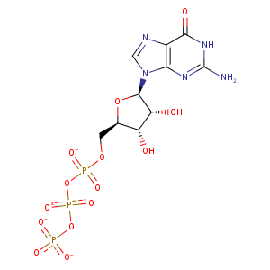
| HET Code: | GTP |
|---|---|
| Formula: | C10H12N5O14P3 |
| Molecular weight: | 519.149 g/mol |
| DrugBank ID: | DB04137 |
| Buried Surface Area: | 63 % |
| Polar Surface area: | 335.56 Å2 |
| Number of | |
|---|---|
| H-Bond Acceptors: | 17 |
| H-Bond Donors: | 4 |
| Rings: | 3 |
| Aromatic rings: | 1 |
| Anionic atoms: | 4 |
| Cationic atoms: | 0 |
| Rule of Five Violation: | 2 |
| Rotatable Bonds: | 8 |
| X | Y | Z |
|---|---|---|
| 55.3206 | 18.2026 | 38.5842 |


Represent the protein/ligand binding mode, centered on the ligand
Dashed lines represents hydrogen bonds and metal interactions
Green residue labels for amino acids with hydrophobic contacts (green lines) to the ligand
| Ligand | Protein | Interaction | |||
|---|---|---|---|---|---|
| Atom | Atom | Residue | Distance (Å) | Angle (°) | Type |
| O1B | N | GLY- 47 | 2.89 | 172.87 | H-Bond (Protein Donor) |
| O2A | N | ALA- 48 | 2.72 | 171.45 | H-Bond (Protein Donor) |
| C1' | CB | ALA- 48 | 4.2 | 0 | Hydrophobic |
| O3G | N | ALA- 97 | 2.73 | 159.88 | H-Bond (Protein Donor) |
| O1G | N | GLY- 99 | 2.6 | 143.5 | H-Bond (Protein Donor) |
| O1G | N | GLY- 134 | 2.84 | 157.87 | H-Bond (Protein Donor) |
| O3G | OG1 | THR- 135 | 2.76 | 175.09 | H-Bond (Protein Donor) |
| O3G | N | THR- 135 | 3.5 | 127.53 | H-Bond (Protein Donor) |
| O3B | N | THR- 135 | 3.06 | 168.07 | H-Bond (Protein Donor) |
| O2B | N | GLY- 136 | 2.68 | 165.96 | H-Bond (Protein Donor) |
| O3' | OE1 | GLU- 165 | 2.63 | 160.21 | H-Bond (Ligand Donor) |
| O2' | OE2 | GLU- 165 | 2.65 | 156.36 | H-Bond (Ligand Donor) |
| C3' | CD | ARG- 169 | 4.33 | 0 | Hydrophobic |
| O3' | NH1 | ARG- 169 | 3.04 | 173.15 | H-Bond (Protein Donor) |
| C2' | CE1 | PHE- 208 | 3.57 | 0 | Hydrophobic |
| N1 | OD1 | ASP- 212 | 2.58 | 141.97 | H-Bond (Ligand Donor) |
| N2 | OD1 | ASP- 212 | 2.59 | 142.06 | H-Bond (Ligand Donor) |
| O2A | O | HOH- 2005 | 2.79 | 179.96 | H-Bond (Protein Donor) |
| O2G | O | HOH- 2035 | 2.94 | 161.85 | H-Bond (Protein Donor) |
| O2B | O | HOH- 2048 | 2.71 | 179.99 | H-Bond (Protein Donor) |
