sc-PDB
An Annotated Database of Druggable Binding Sites from the Protein DataBank
An Annotated Database of Druggable Binding Sites from the Protein DataBank
2.000 Å
X-ray
2003-10-20
| Name: | Trehalose-6-phosphate synthase |
|---|---|
| ID: | OTSA_ECOLI |
| AC: | P31677 |
| Organism: | Escherichia coli |
| Reign: | Bacteria |
| TaxID: | 83333 |
| EC Number: | 2.4.1.15 |
| Chain Name: | Percentage of Residues within binding site |
|---|---|
| A | 100 % |
| B-Factor: | 19.821 |
|---|---|
| Number of residues: | 44 |
| Including | |
| Standard Amino Acids: | 43 |
| Non Standard Amino Acids: | 0 |
| Water Molecules: | 1 |
| Cofactors: | |
| Metals: | |
| Ligandability | Volume (Å3) |
|---|---|
| 0.805 | 918.000 |
| % Hydrophobic | % Polar |
|---|---|
| 44.85 | 55.15 |
| According to VolSite | |
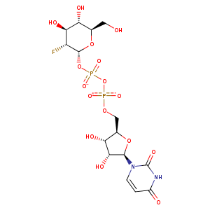
| HET Code: | U2F |
|---|---|
| Formula: | C15H21FN2O16P2 |
| Molecular weight: | 566.277 g/mol |
| DrugBank ID: | DB03488 |
| Buried Surface Area: | 65.31 % |
| Polar Surface area: | 296.59 Å2 |
| Number of | |
|---|---|
| H-Bond Acceptors: | 16 |
| H-Bond Donors: | 6 |
| Rings: | 3 |
| Aromatic rings: | 0 |
| Anionic atoms: | 2 |
| Cationic atoms: | 0 |
| Rule of Five Violation: | 3 |
| Rotatable Bonds: | 9 |
| X | Y | Z |
|---|---|---|
| 31.5001 | 24.3816 | 15.0606 |
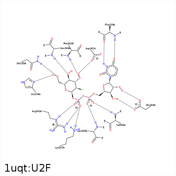


Represent the protein/ligand binding mode, centered on the ligand
Dashed lines represents hydrogen bonds and metal interactions
Green residue labels for amino acids with hydrophobic contacts (green lines) to the ligand
| Ligand | Protein | Interaction | |||
|---|---|---|---|---|---|
| Atom | Atom | Residue | Distance (Å) | Angle (°) | Type |
| F1 | CH2 | TRP- 85 | 3.63 | 0 | Hydrophobic |
| C6 | CB | HIS- 154 | 4.32 | 0 | Hydrophobic |
| O6 | ND1 | HIS- 154 | 2.68 | 152.08 | H-Bond (Ligand Donor) |
| F1 | CG2 | ILE- 155 | 4.11 | 0 | Hydrophobic |
| O6 | NE2 | GLN- 185 | 2.84 | 131.96 | H-Bond (Protein Donor) |
| C6 | CG2 | ILE- 225 | 3.92 | 0 | Hydrophobic |
| O1B | NE | ARG- 262 | 2.74 | 161.76 | H-Bond (Protein Donor) |
| O2B | NH2 | ARG- 262 | 2.9 | 177.11 | H-Bond (Protein Donor) |
| O1B | CZ | ARG- 262 | 3.6 | 0 | Ionic (Protein Cationic) |
| O2B | CZ | ARG- 262 | 3.74 | 0 | Ionic (Protein Cationic) |
| O2B | NZ | LYS- 267 | 3.16 | 166.61 | H-Bond (Protein Donor) |
| O3A | NZ | LYS- 267 | 3.16 | 127.7 | H-Bond (Protein Donor) |
| O2B | NZ | LYS- 267 | 3.16 | 0 | Ionic (Protein Cationic) |
| N3 | O | PHE- 339 | 2.75 | 156.76 | H-Bond (Ligand Donor) |
| O7' | N | PHE- 339 | 3.08 | 168.28 | H-Bond (Protein Donor) |
| O3' | NE | ARG- 341 | 3.42 | 123.68 | H-Bond (Protein Donor) |
| O3 | OD1 | ASP- 361 | 2.57 | 146.39 | H-Bond (Ligand Donor) |
| O3 | N | MET- 363 | 2.82 | 174.09 | H-Bond (Protein Donor) |
| C4 | CB | MET- 363 | 4.15 | 0 | Hydrophobic |
| O3 | N | ASN- 364 | 3.24 | 121.04 | H-Bond (Protein Donor) |
| O4 | N | ASN- 364 | 2.81 | 153.84 | H-Bond (Protein Donor) |
| O1A | N | LEU- 365 | 2.98 | 174.55 | H-Bond (Protein Donor) |
| C6 | CD1 | LEU- 365 | 4.22 | 0 | Hydrophobic |
| O2A | N | VAL- 366 | 3.31 | 162.01 | H-Bond (Protein Donor) |
| C5' | CG2 | VAL- 366 | 4.28 | 0 | Hydrophobic |
| C2' | CG2 | VAL- 366 | 3.78 | 0 | Hydrophobic |
| O3' | OE1 | GLU- 369 | 2.53 | 156.9 | H-Bond (Ligand Donor) |
| O2' | OE1 | GLU- 369 | 3.38 | 135.33 | H-Bond (Ligand Donor) |
| O2' | OE2 | GLU- 369 | 2.63 | 163.29 | H-Bond (Ligand Donor) |
