sc-PDB
An Annotated Database of Druggable Binding Sites from the Protein DataBank
An Annotated Database of Druggable Binding Sites from the Protein DataBank
2.000 Å
X-ray
2004-08-04
| Name: | 3-hydroxyacyl-CoA dehydrogenase type-2 |
|---|---|
| ID: | HCD2_HUMAN |
| AC: | Q99714 |
| Organism: | Homo sapiens |
| Reign: | Eukaryota |
| TaxID: | 9606 |
| EC Number: | 1.1.1.35 |
| Chain Name: | Percentage of Residues within binding site |
|---|---|
| A | 100 % |
| B-Factor: | 25.238 |
|---|---|
| Number of residues: | 65 |
| Including | |
| Standard Amino Acids: | 61 |
| Non Standard Amino Acids: | 0 |
| Water Molecules: | 4 |
| Cofactors: | |
| Metals: | |
| Ligandability | Volume (Å3) |
|---|---|
| 1.436 | 961.875 |
| % Hydrophobic | % Polar |
|---|---|
| 50.88 | 49.12 |
| According to VolSite | |

| HET Code: | TDT |
|---|---|
| Formula: | C40H46N12O15P2S |
| Molecular weight: | 1028.877 g/mol |
| DrugBank ID: | DB02820 |
| Buried Surface Area: | 68.39 % |
| Polar Surface area: | 428.72 Å2 |
| Number of | |
|---|---|
| H-Bond Acceptors: | 23 |
| H-Bond Donors: | 6 |
| Rings: | 9 |
| Aromatic rings: | 4 |
| Anionic atoms: | 2 |
| Cationic atoms: | 0 |
| Rule of Five Violation: | 3 |
| Rotatable Bonds: | 15 |
| X | Y | Z |
|---|---|---|
| 81.009 | 8.87254 | 35.7815 |
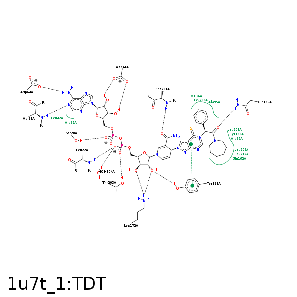


Represent the protein/ligand binding mode, centered on the ligand
Dashed lines represents hydrogen bonds and metal interactions
Green residue labels for amino acids with hydrophobic contacts (green lines) to the ligand
| Ligand | Protein | Interaction | |||
|---|---|---|---|---|---|
| Atom | Atom | Residue | Distance (Å) | Angle (°) | Type |
| O2A | OG | SER- 20 | 2.51 | 152.97 | H-Bond (Protein Donor) |
| C3B | CB | SER- 20 | 4.06 | 0 | Hydrophobic |
| O2N | N | LEU- 22 | 2.88 | 161.62 | H-Bond (Protein Donor) |
| C5M | CD1 | LEU- 22 | 4.27 | 0 | Hydrophobic |
| O3B | OD1 | ASP- 41 | 3.17 | 133.58 | H-Bond (Ligand Donor) |
| O2B | OD1 | ASP- 41 | 2.66 | 165.75 | H-Bond (Ligand Donor) |
| N6A | OD2 | ASP- 64 | 2.66 | 174.22 | H-Bond (Ligand Donor) |
| N1A | N | VAL- 65 | 2.82 | 164.38 | H-Bond (Protein Donor) |
| C5M | CB | CYS- 91 | 4.05 | 0 | Hydrophobic |
| C1B | CB | ALA- 92 | 4.35 | 0 | Hydrophobic |
| C10 | CB | ALA- 95 | 3.92 | 0 | Hydrophobic |
| C13 | CB | ALA- 97 | 3.58 | 0 | Hydrophobic |
| C4M | CG2 | THR- 153 | 4.02 | 0 | Hydrophobic |
| C1M | CG2 | THR- 153 | 3.75 | 0 | Hydrophobic |
| C5 | CB | GLN- 162 | 3.66 | 0 | Hydrophobic |
| O | NE2 | GLN- 165 | 2.54 | 137.43 | H-Bond (Protein Donor) |
| C5 | CG | GLN- 165 | 4 | 0 | Hydrophobic |
| O2M | OH | TYR- 168 | 2.61 | 156.64 | H-Bond (Protein Donor) |
| O3M | NZ | LYS- 172 | 2.88 | 130.67 | H-Bond (Protein Donor) |
| O2M | NZ | LYS- 172 | 3 | 143.59 | H-Bond (Protein Donor) |
| C4N | CB | PRO- 198 | 4.27 | 0 | Hydrophobic |
| O7N | N | PHE- 201 | 2.59 | 162.9 | H-Bond (Protein Donor) |
| N7N | O | PHE- 201 | 3.4 | 143.91 | H-Bond (Ligand Donor) |
| O1N | OG1 | THR- 203 | 2.58 | 170.54 | H-Bond (Protein Donor) |
| C2M | CD2 | LEU- 205 | 4.17 | 0 | Hydrophobic |
| C | CD1 | LEU- 209 | 4.3 | 0 | Hydrophobic |
| C3 | CD1 | LEU- 209 | 3.78 | 0 | Hydrophobic |
| C8 | CD2 | LEU- 209 | 4.23 | 0 | Hydrophobic |
| C9 | CD1 | LEU- 209 | 3.83 | 0 | Hydrophobic |
| C10 | CG | LEU- 209 | 4.09 | 0 | Hydrophobic |
| C11 | CD2 | LEU- 209 | 4.44 | 0 | Hydrophobic |
| C3 | CG1 | VAL- 213 | 3.98 | 0 | Hydrophobic |
| C7 | CD1 | LEU- 217 | 4.01 | 0 | Hydrophobic |
| O2N | O | HOH- 504 | 2.98 | 179.97 | H-Bond (Protein Donor) |
